Anterolisthesis at the cervicothoracic junction represents one of the most complex spinal conditions affecting the critical transition zone between the highly mobile cervical spine and the relatively rigid thoracic region. When the seventh cervical vertebra (C7) slips forward over the first thoracic vertebra (T1), it creates a unique biomechanical challenge that can significantly impact both structural stability and neurological function. This condition occurs at a junction where the natural lordotic curve of the neck transitions into the kyphotic curve of the upper back, creating inherent stress concentrations that predispose this region to instability.
The cervicothoracic junction serves as a crucial anatomical crossroads where multiple biomechanical forces converge. Unlike anterolisthesis in the lumbar spine, which is more commonly recognised and studied, C7-T1 anterolisthesis presents unique diagnostic and therapeutic challenges due to the complex anatomy of this transitional zone. The condition affects approximately 2-9% of all cervical spine disorders, yet it remains one of the most frequently missed diagnoses in emergency settings due to imaging difficulties and the subtle nature of early symptoms.
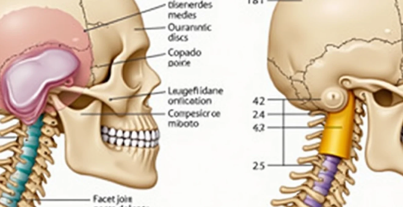
Cervicothoracic junction anatomy and C7-T1 vertebral alignment
The cervicothoracic junction represents a remarkable anatomical transition where the mobile cervical spine meets the stabilised thoracic region. This junction experiences approximately half the flexibility of the upper cervical segments, creating a natural fulcrum point where mechanical stresses concentrate during movement. The anatomical complexity of this region stems from the dramatic change in spinal curvature, vertebral morphology, and the presence of the first rib articulation, all of which contribute to the unique biomechanical environment that can predispose to anterolisthesis.
Anatomical structure of the seventh cervical vertebra
The seventh cervical vertebra, commonly known as vertebra prominens , exhibits distinctive anatomical features that bridge the cervical and thoracic regions. Its elongated spinous process extends prominently beneath the skin at the base of the neck, serving as a crucial anatomical landmark for clinical examination. The vertebral body of C7 is larger and more robust than other cervical vertebrae, reflecting its role in supporting increased mechanical loads at the cervicothoracic junction. The transverse processes lack the characteristic foramen transversarium found in other cervical vertebrae, indicating the absence of vertebral artery passage at this level.
First thoracic vertebra morphological characteristics
The first thoracic vertebra marks the beginning of the thoracic spine and demonstrates unique morphological adaptations for rib articulation. T1 features complete costal facets on its vertebral body for articulation with the first rib, creating additional stability but also introducing new stress patterns. The vertebral body dimensions are intermediate between cervical and thoracic proportions, whilst the spinous process projects at a more acute downward angle compared to C7. These anatomical variations contribute to the complex biomechanical environment where anterolisthesis may develop, as the vertebra must accommodate both spinal column loading and rib cage dynamics.
Intervertebral disc composition at C7-T1 level
The C7-T1 intervertebral disc demonstrates unique compositional characteristics that reflect the transitional nature of this spinal segment. The annulus fibrosus exhibits a more robust construction with increased collagen density compared to upper cervical discs, whilst maintaining the flexibility required for cervical motion. The nucleus pulposus occupies a relatively smaller proportion of the disc space, reflecting the increased mechanical demands at this level. Degenerative changes in this disc often occur earlier than in other cervical segments due to the concentrated stress patterns, making it a frequent site for disc-related anterolisthesis.
Facet joint orientation and biomechanical function
The facet joints at the C7-T1 level exhibit a transitional orientation that bridges the more horizontal cervical facets with the vertical thoracic configuration. This intermediate angulation, typically ranging between 45-60 degrees from the horizontal plane, creates unique loading patterns during cervical motion. The facet joint capsules at this level are particularly susceptible to degenerative changes due to the concentrated rotational and translational forces that occur during normal cervical spine movement. These biomechanical factors contribute significantly to the development of facet joint-mediated anterolisthesis at the cervicothoracic junction.
Pathophysiology of anterolisthesis at the cervicothoracic junction
The development of anterolisthesis at the C7-T1 level involves complex pathophysiological mechanisms that reflect the unique biomechanical environment of the cervicothoracic junction. Multiple factors contribute to the forward displacement of C7 on T1, including degenerative processes, traumatic injuries, and congenital abnormalities. The pathophysiology differs significantly from lumbar anterolisthesis due to the distinct anatomical characteristics and loading patterns of the cervical spine. Understanding these mechanisms is crucial for developing effective treatment strategies and preventing progression of the condition.
Degenerative disc disease contributing to vertebral slippage
Degenerative disc disease at the C7-T1 level represents a primary pathological mechanism leading to anterolisthesis development. The intervertebral disc experiences accelerated degeneration due to the concentrated mechanical stresses at the cervicothoracic junction. As the nucleus pulposus loses hydration and the annulus fibrosus develops fissures, the disc’s ability to maintain vertebral alignment diminishes progressively. This process typically begins in the third decade of life but may accelerate due to repetitive trauma, genetic predisposition, or metabolic factors. The resulting disc height loss and altered load distribution create conditions favourable for anterior vertebral translation.
Ligamentous instability patterns in C7-T1 anterolisthesis
Ligamentous structures play a critical role in maintaining C7-T1 stability, and their compromise contributes significantly to anterolisthesis development. The posterior longitudinal ligament, ligamentum flavum, and interspinous ligaments work synergistically to resist anterior translation of C7. Age-related degeneration, traumatic injury, or inflammatory processes can weaken these structures, leading to progressive instability. The ligamentous laxity often develops insidiously, with patients initially experiencing intermittent symptoms that gradually become more persistent as the instability progresses. Imaging studies frequently reveal thickening or discontinuity of these ligamentous structures in patients with established anterolisthesis.
Facet joint degeneration and capsular laxity
Facet joint pathology represents another crucial component in the pathogenesis of C7-T1 anterolisthesis. The unique orientation and loading patterns of the cervicothoracic facet joints predispose them to degenerative changes, including cartilage loss, subchondral sclerosis, and osteophyte formation. Capsular laxity develops as a consequence of chronic inflammation and repetitive microtrauma, reducing the joint’s ability to resist translational forces. This process is often accelerated by occupational factors, such as prolonged computer work or repetitive overhead activities, which place sustained stress on the cervicothoracic junction.
Biomechanical forces during cervical Flexion-Extension
The cervicothoracic junction experiences complex biomechanical forces during normal cervical spine movement, particularly during flexion-extension cycles. Forward flexion creates anterior compression forces and posterior tension, whilst the rigid thoracic spine restricts motion below T1, concentrating stress at the C7-T1 level. Extension movements generate anterior tension and posterior compression, with the potential for facet joint overloading. These repetitive stress patterns, combined with age-related tissue changes, can gradually overwhelm the stabilising structures and lead to progressive anterior slippage. Whiplash injuries particularly exploit these biomechanical vulnerabilities, potentially causing acute or delayed anterolisthesis development.
Clinical manifestations and neurological implications
The clinical presentation of C7-T1 anterolisthesis varies considerably depending on the degree of vertebral displacement, associated neural compression, and individual patient factors. Unlike lumbar anterolisthesis, which primarily affects the lower extremities, cervicothoracic anterolisthesis can produce a complex array of symptoms affecting the neck, shoulders, arms, and even the upper back. The proximity to vital neurological structures, including the C8 nerve root and the cervical spinal cord, makes this condition particularly significant from a clinical perspective.
Patients typically present with progressive neck pain that may radiate into the shoulders and upper extremities. The pain often demonstrates a characteristic pattern, worsening with neck extension and improving with flexion, reflecting the biomechanical nature of the underlying instability. Neurological symptoms may include numbness, tingling, or weakness in the C8 dermatome distribution, affecting the medial forearm, hand, and little finger. In severe cases, patients may experience difficulty with fine motor tasks, such as writing or buttoning clothes, due to intrinsic hand muscle weakness.
The neurological implications extend beyond simple nerve root compression. Spinal cord compromise, whilst less common, can occur in high-grade anterolisthesis and may result in myelopathic symptoms including gait disturbance, upper extremity clumsiness, and bladder dysfunction. Cervical myelopathy associated with C7-T1 anterolisthesis requires urgent attention, as progressive cord compression can lead to irreversible neurological deficits. Patients may also experience referred pain patterns, with symptoms extending into the occipital region, temporal area, or even the anterior chest wall due to the complex innervation patterns of the cervicothoracic region.
The clinical challenge lies in recognising the often subtle early symptoms of C7-T1 anterolisthesis, which may masquerade as common conditions such as tension headaches, shoulder impingement, or carpal tunnel syndrome.
Diagnostic imaging protocols for C7-T1 anterolisthesis
Accurate diagnosis of C7-T1 anterolisthesis requires a systematic approach utilising multiple imaging modalities. The cervicothoracic junction presents unique imaging challenges due to overlapping anatomical structures, including the shoulder girdle, first ribs, and lung apices. Standard cervical spine radiographs often fail to adequately visualise this region, necessitating specialised imaging techniques and positioning to achieve diagnostic quality images.
Lateral cervical radiograph measurement techniques
Lateral cervical radiographs remain the primary screening tool for detecting C7-T1 anterolisthesis, though technical factors significantly influence image quality at the cervicothoracic junction. Proper positioning requires shoulder depression, often achieved through gentle traction or patient positioning modifications. The percentage slip calculation follows established protocols, measuring the anterior displacement of the C7 vertebral body relative to the superior endplate of T1. Additional measurements include assessment of the cervicothoracic angle and evaluation of spinous process alignment, which can provide early indicators of instability even before obvious vertebral displacement occurs.
MRI T1 and T2 weighted sequence analysis
Magnetic resonance imaging provides superior soft tissue contrast and detailed visualisation of neural structures affected by C7-T1 anterolisthesis. T2-weighted sagittal sequences excel at demonstrating disc degeneration, ligamentous injury, and spinal cord compression, whilst T1-weighted images provide excellent anatomical detail and can identify bone marrow changes associated with chronic instability. STIR (Short Tau Inversion Recovery) sequences prove particularly valuable for detecting bone marrow oedema and ligamentous inflammation. The addition of gradient echo sequences enhances visualisation of cerebrospinal fluid flow dynamics and can identify subtle spinal cord compression that might not be apparent on conventional sequences.
CT myelography for spinal canal assessment
CT myelography remains the gold standard for detailed evaluation of spinal canal dimensions and neural element compression in C7-T1 anterolisthesis. This technique provides superior bony detail compared to MRI and can accurately assess the degree of canal stenosis, particularly in patients with significant degenerative changes or previous surgical intervention. The three-dimensional reconstruction capabilities of modern CT systems allow for detailed assessment of facet joint orientation and identification of bony abnormalities that may contribute to instability. Intrathecal contrast administration enables precise localisation of nerve root compression and can guide surgical planning when conservative management fails.
Dynamic Flexion-Extension radiographic studies
Dynamic imaging studies play a crucial role in assessing the functional significance of C7-T1 anterolisthesis and determining treatment recommendations. Flexion-extension radiographs can demonstrate abnormal motion patterns, quantify translational instability, and identify patients at risk for progression. The technique requires careful patient cooperation and radiographer expertise to ensure adequate flexion and extension whilst maintaining patient safety. Measurements typically focus on translational motion exceeding 3.5mm or angular motion greater than 11 degrees between adjacent vertebrae. These studies prove particularly valuable in differentiating between stable degenerative changes and clinically significant instability requiring intervention.
Meyerding classification system applied to cervical anterolisthesis
The Meyerding classification system, originally developed for lumbar spondylolisthesis, has been adapted for use in cervical anterolisthesis, including C7-T1 displacement. This grading system provides a standardised method for quantifying the degree of vertebral slippage and guides treatment decision-making. The classification divides the superior endplate of the lower vertebra into four equal quarters, with the degree of forward displacement determining the grade. Grade I represents 0-25% displacement, Grade II indicates 25-50% slip, Grade III encompasses 50-75% displacement, and Grade IV represents 75-100% anterior translation.
Application of the Meyerding system to cervical anterolisthesis requires careful consideration of the unique anatomical characteristics of the cervical spine. The smaller vertebral body dimensions and different loading patterns compared to the lumbar spine influence both the measurement technique and clinical significance of each grade. Low-grade slips (Grades I and II) in the cervical spine may produce more significant symptoms than comparable lumbar slips due to the proximity of neural structures and the smaller spinal canal dimensions. High-grade cervical anterolisthesis (Grades III and IV) represents a surgical emergency due to the high risk of spinal cord injury.
The classification system serves multiple purposes in clinical practice, including standardising communication between healthcare providers, tracking progression over time, and determining appropriate treatment strategies. Grade I and II slips typically respond well to conservative management, including physical therapy, activity modification, and anti-inflammatory medications. Grade III slips often require more aggressive intervention, with surgical stabilisation frequently necessary to prevent progression and neurological compromise. Grade IV anterolisthesis almost universally requires surgical treatment due to the significant risk of complete spinal cord injury.
The Meyerding classification system provides crucial prognostic information, with higher grades correlating with increased risk of neurological complications and reduced success rates with conservative treatment approaches.
Conservative treatment modalities and surgical interventions
Treatment of C7-T1 anterolisthesis follows a graduated approach, with conservative management typically attempted first for low-grade slips without significant neurological compromise. The unique biomechanical environment of the cervicothoracic junction influences treatment selection, with particular attention paid to maintaining cervical spine mobility whilst addressing instability. Conservative treatment modalities include activity modification, pharmacological management, physical therapy, and orthotic support, each tailored to the individual patient’s presentation and functional requirements.
Physical therapy plays a central role in conservative management, focusing on strengthening the deep cervical flexors, improving posture, and enhancing cervicothoracic stability. Isometric strengthening exercises prove particularly beneficial, as they improve muscle endurance without placing excessive stress on the unstable segment. Manual therapy techniques, including gentle mobilisation and soft tissue techniques, can address associated muscle tension and improve overall function. Patient education regarding proper ergonomics and activity modification helps prevent symptom exacerbation and may slow progression of the condition.
Pharmacological interventions typically begin with anti-inflammatory medications, including NSAIDs and topical preparations, which address the inflammatory component of the condition. Muscle relaxants may provide short-term relief for associated cervical muscle spasm, whilst neuropathic pain medications can be beneficial for patients experiencing radicular symptoms. Epidural steroid injections, whilst technically challenging at the cervicothoracic junction, may provide temporary relief and help identify patients who might benefit from surgical
intervention.
Cervical orthoses may provide external support for patients with mild to moderate instability, though their effectiveness at the cervicothoracic junction remains limited due to the anatomical challenges of immobilising this region. Soft collars offer minimal restriction but may provide proprioceptive feedback and psychological comfort. Rigid cervical orthoses, such as Philadelphia or Miami J collars, provide greater immobilisation but can be poorly tolerated due to their bulk and restriction of daily activities. The decision to use orthotic support must balance the potential benefits against the risk of muscle deconditioning and psychological dependence.
Surgical intervention becomes necessary when conservative treatment fails, neurological symptoms progress, or high-grade instability threatens spinal cord integrity. The surgical approach to C7-T1 anterolisthesis requires careful consideration of the unique anatomical challenges presented by the cervicothoracic junction. Posterior cervical fusion remains the most commonly employed technique, utilising lateral mass screws in C7 and pedicle or pars screws in T1 to achieve rigid fixation. The procedure typically involves decompression of neural elements followed by instrumented fusion, with autograft or allograft bone placement to promote solid arthrodesis.
Anterior cervical approaches present technical challenges at the C7-T1 level but may be considered in cases where anterior pathology predominates or when posterior approaches are contraindicated. The proximity of vascular structures, including the vertebral artery and carotid vessels, requires meticulous surgical technique and thorough preoperative planning. Combined anterior-posterior approaches may be necessary for complex cases involving significant bone destruction, tumour involvement, or severe deformity correction requirements.
The success of surgical intervention depends on multiple factors, including patient age, bone quality, smoking status, and the degree of preoperative neurological compromise. Fusion rates at the cervicothoracic junction typically range from 85-95% with modern instrumentation techniques, though the healing process may be prolonged compared to other cervical levels due to the unique biomechanical stresses. Postoperative rehabilitation focuses on gradual return to activity whilst protecting the fusion site during the initial healing phase, typically requiring 3-6 months for solid arthrodesis to develop.
What factors influence your choice between conservative management and surgical intervention for C7-T1 anterolisthesis? The decision-making process requires careful assessment of symptom severity, neurological involvement, patient age and activity level, and response to initial conservative measures. Patients with progressive neurological deficits, high-grade instability, or failure of comprehensive conservative treatment over 6-12 months typically warrant surgical consideration. The goal of any intervention, whether conservative or surgical, remains the same: to relieve symptoms, prevent neurological deterioration, and restore functional capacity whilst minimising treatment-related morbidity.
Long-term outcomes following treatment of C7-T1 anterolisthesis vary considerably based on the chosen intervention and patient factors. Conservative treatment success rates range from 60-80% for low-grade slips, with most patients experiencing significant symptom improvement within 3-6 months of initiating treatment. Surgical outcomes demonstrate excellent results in appropriately selected patients, with most studies reporting good to excellent outcomes in 80-90% of cases. However, the complex nature of cervicothoracic junction pathology means that some patients may experience persistent symptoms despite technically successful treatment, emphasising the importance of comprehensive preoperative counselling and realistic expectation setting.
The key to successful management of C7-T1 anterolisthesis lies in early recognition, appropriate classification, and timely intervention tailored to the individual patient’s presentation and functional requirements.
Understanding anterolisthesis of C7 on T1 requires appreciation of the complex anatomical and biomechanical factors that make the cervicothoracic junction vulnerable to instability. The condition presents unique diagnostic and therapeutic challenges that differ significantly from more commonly encountered lumbar spondylolisthesis. Through careful application of diagnostic imaging protocols, appropriate classification systems, and evidence-based treatment approaches, healthcare providers can achieve optimal outcomes for patients affected by this challenging condition. The evolution of surgical techniques and conservative management strategies continues to improve prognosis, though the fundamental principles of early recognition and individualised treatment remain paramount to successful management.