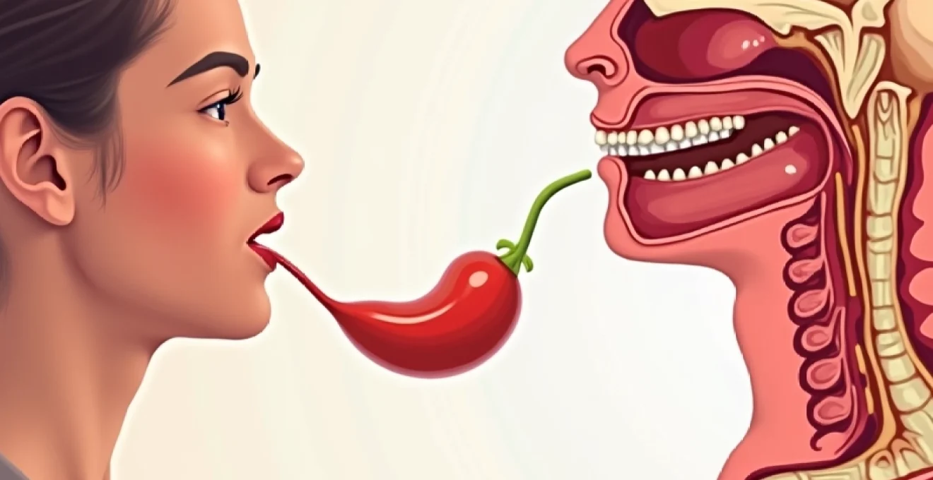
Thyroid disorders affect millions of people worldwide, yet the connection between these endocrine conditions and swallowing problems remains poorly understood by many patients and healthcare providers alike. When the thyroid gland malfunctions, it can create a cascade of effects that significantly impact the delicate mechanisms involved in deglutition. From mechanical compression caused by enlarged thyroid tissue to neurological complications affecting the muscles and nerves essential for swallowing, thyroid pathology presents a complex array of challenges that can profoundly affect quality of life and nutritional status.
The relationship between thyroid dysfunction and dysphagia is multifaceted, involving both direct anatomical interference and indirect metabolic effects on muscle function and tissue composition. Understanding these connections is crucial for both patients experiencing unexplained swallowing difficulties and healthcare professionals seeking to provide comprehensive care for thyroid-related complications.
Thyroid anatomy and its direct impact on swallowing mechanisms
The thyroid gland’s strategic location in the anterior neck places it in intimate proximity to the structures essential for normal swallowing function. This butterfly-shaped endocrine organ sits directly adjacent to the trachea, oesophagus, and the complex network of muscles and nerves that coordinate the swallowing process. When thyroid pathology develops, whether through enlargement, inflammation, or malignancy, the resulting anatomical changes can create significant mechanical interference with deglutition.
Thyroid gland positioning adjacent to oesophageal and tracheal structures
The thyroid gland’s anatomical relationship with the oesophagus and trachea creates a scenario where even modest enlargement can produce compressive symptoms. Research indicates that up to 33% of patients with benign goitres experience difficulty swallowing , highlighting the prevalence of this complication. The oesophagus passes directly behind the thyroid gland, separated only by a thin layer of fascial tissue and the trachea itself.
When thyroid enlargement occurs, the expanding tissue can compress the oesophagus against the vertebral column, creating a mechanical obstruction to food passage. This compression typically affects the cervical portion of the oesophagus most significantly, as this region has the least room for accommodation. Patients often describe a sensation of food becoming lodged in the throat or chest, particularly with solid foods that require more space for passage.
The degree of compression varies considerably depending on the size, location, and growth pattern of the thyroid enlargement. Substernal goitres, which extend below the clavicle into the mediastinum, can create particularly severe compressive symptoms as they grow within the confined space of the thoracic inlet. These cases often require surgical intervention even when the thyroid tissue is histologically benign.
Recurrent laryngeal nerve compression in thyroid enlargement
The recurrent laryngeal nerves play a crucial role in vocal cord function and contribute to the protective mechanisms during swallowing. These delicate neural structures course through the tracheoesophageal groove, positioned directly adjacent to the thyroid gland’s posterior aspect. When thyroid pathology develops, particularly with posterior extension of nodules or inflammatory processes, these nerves can become compressed or stretched.
Recurrent laryngeal nerve dysfunction can manifest as hoarseness, aspiration risk, and altered swallowing coordination . The nerve’s role in vocal cord adduction becomes compromised, leading to incomplete glottic closure during swallowing. This incomplete closure allows food particles and liquids to penetrate the laryngeal vestibule, increasing the risk of aspiration pneumonia.
The vulnerability of the recurrent laryngeal nerve to thyroid-related compression varies between individuals based on anatomical variations and the specific pattern of thyroid enlargement. Malignant thyroid processes pose a higher risk for nerve involvement due to their infiltrative growth pattern, but benign conditions can also cause significant neural compromise through gradual compression over time.
Cricoarytenoid muscle dysfunction from thyroid pathology
The cricoarytenoid muscles, innervated by the recurrent laryngeal nerves, are essential for proper laryngeal function during swallowing. When thyroid disease affects the neural supply to these muscles, their ability to coordinate vocal cord movement becomes impaired. This impairment particularly affects the posterior cricoarytenoid muscles, which are responsible for vocal cord abduction and play a protective role during respiration.
Cricoarytenoid muscle dysfunction can lead to paradoxical vocal cord movement during swallowing, where the cords fail to adduct properly for airway protection or fail to abduct adequately for breathing. Patients may experience a chronic cough, throat clearing, or a sensation of incomplete swallowing. The muscle weakness progresses gradually in most cases, allowing for some compensation, but acute changes can occur with rapid thyroid enlargement or inflammatory processes.
Superior laryngeal nerve involvement in thyroid disease
While less commonly affected than the recurrent laryngeal nerve, the superior laryngeal nerve can also be compromised by thyroid pathology. This nerve provides sensory innervation to the laryngeal inlet and motor innervation to the cricothyroid muscle. When affected, patients may experience altered sensation in the throat, affecting their ability to detect food or liquid approaching the larynx.
Superior laryngeal nerve dysfunction particularly impacts the timing of swallowing reflexes. The nerve’s sensory component triggers the pharyngeal phase of swallowing, and when this trigger mechanism is compromised, patients may experience delayed or absent swallowing reflexes. This delay increases the risk of aspiration as food or liquid may enter the airway before protective mechanisms activate.
Hypothyroidism-related dysphagia and myxoedema effects
Hypothyroidism creates a unique set of swallowing challenges that extend beyond simple mechanical compression. The metabolic effects of thyroid hormone deficiency profoundly impact muscle function, tissue composition, and neural transmission throughout the body, including the structures involved in deglutition. These systemic effects can produce swallowing difficulties even in the absence of thyroid gland enlargement.
Macroglossia development in severe hypothyroidism
Severe hypothyroidism can lead to macroglossia, an enlargement of the tongue that significantly impacts swallowing function. This enlargement results from myxoedematous infiltration of the tongue muscles , where glycosaminoglycans accumulate in the tissue spaces, causing swelling and reduced muscle efficiency. The enlarged tongue occupies more space within the oral cavity, interfering with the oral preparatory phase of swallowing.
Macroglossia affects multiple aspects of deglutition, from initial food manipulation to the propulsion of the food bolus into the pharynx. Patients may notice difficulty chewing, problems with speech articulation, and a sensation that their tongue feels too large for their mouth. The condition typically develops gradually over months or years, allowing patients to adapt somewhat to the changes, but severe cases can create significant functional impairment.
The degree of macroglossia correlates with the severity and duration of hypothyroidism. Early recognition and treatment with thyroid hormone replacement can lead to gradual improvement in tongue size and function, though complete resolution may take several months of adequate treatment. In some cases, residual effects may persist even after achieving euthyroid status.
Oesophageal motility disorders in hashimoto’s thyroiditis
Hashimoto’s thyroiditis, the most common cause of hypothyroidism in iodine-sufficient areas, can be associated with oesophageal motility disorders that extend beyond the effects of simple hormone deficiency. The autoimmune process underlying Hashimoto’s thyroiditis may affect the smooth muscle of the oesophagus directly, leading to impaired peristaltic function.
Studies have shown that patients with Hashimoto’s thyroiditis may develop oesophageal dysmotility even before developing overt hypothyroidism, suggesting a direct immune-mediated effect on oesophageal smooth muscle.
The oesophageal motility changes in Hashimoto’s thyroiditis typically manifest as weakened peristaltic waves, reduced lower oesophageal sphincter pressure, and delayed oesophageal transit times. These changes can lead to symptoms of gastroesophageal reflux, food stasis in the oesophagus, and a sensation of incomplete swallowing. The motility disorders may improve with thyroid hormone replacement therapy, but some patients require additional treatment for persistent symptoms.
Pharyngeal muscle weakness from thyroid hormone deficiency
Thyroid hormone deficiency affects skeletal muscle function throughout the body, including the muscles responsible for pharyngeal contraction during swallowing. The pharyngeal muscles require coordinated, forceful contraction to propel the food bolus from the mouth into the oesophagus whilst simultaneously protecting the airway. When thyroid hormone levels are insufficient, these muscles may become weak and poorly coordinated.
Pharyngeal muscle weakness in hypothyroidism manifests as reduced pharyngeal constriction strength and delayed pharyngeal transit times. Patients may experience food sticking in the throat, require multiple swallows to clear food from the pharynx, or develop a sensation of incomplete swallowing. The weakness typically affects all pharyngeal muscles uniformly, rather than showing the asymmetric patterns seen with neurological disorders.
The muscle weakness responds to thyroid hormone replacement therapy, but improvement may be gradual and incomplete in patients with longstanding hypothyroidism. Some degree of muscle fibre damage may occur with prolonged hormone deficiency, leading to persistent weakness even after achieving normal thyroid function. Early recognition and treatment of hypothyroidism are therefore crucial for preventing irreversible muscle changes.
Myxoedematous infiltration of laryngeal tissues
The accumulation of glycosaminoglycans in laryngeal tissues represents another mechanism by which hypothyroidism can affect swallowing function. This myxoedematous infiltration causes thickening and stiffening of the vocal cords, aryepiglottic folds, and other laryngeal structures essential for airway protection during swallowing.
Laryngeal myxoedema particularly affects the mobility and sensitivity of the epiglottis and vocal cords. The epiglottis may become thickened and less mobile, reducing its effectiveness in covering the laryngeal inlet during swallowing. Similarly, vocal cord thickening can impair their ability to close completely, increasing the risk of penetration and aspiration. Patients often notice voice changes, including hoarseness and reduced vocal range, alongside their swallowing difficulties.
Hyperthyroidism and graves’ disease swallowing complications
Hyperthyroidism presents a different set of challenges for swallowing function, primarily through its effects on muscle metabolism and the potential for significant thyroid enlargement. The hypermetabolic state characteristic of hyperthyroidism can lead to muscle weakness and altered coordination, whilst conditions like Graves’ disease may produce additional complications through extrathyroidal manifestations and diffuse goitre formation.
Thyrotoxic myopathy affecting deglutition muscles
Thyrotoxic myopathy represents a significant complication of hyperthyroidism that can profoundly impact swallowing function. This condition affects both skeletal and smooth muscle throughout the body, including the muscles responsible for deglutition. The excess thyroid hormone accelerates protein catabolism whilst impairing protein synthesis, leading to progressive muscle weakness despite normal or increased appetite.
The deglutition muscles affected by thyrotoxic myopathy include the mylohyoid, geniohyoid, and stylohyoid muscles responsible for laryngeal elevation, as well as the pharyngeal constrictors essential for bolus propulsion. Patients may experience difficulty initiating swallows, reduced pharyngeal pressure generation, and incomplete laryngeal elevation during swallowing. These changes increase the risk of aspiration and can lead to significant nutritional compromise.
The muscle weakness in thyrotoxic myopathy typically develops gradually over weeks to months, paralleling the progression of the hyperthyroid state. Unlike some other causes of muscle weakness, thyrotoxic myopathy generally responds well to treatment of the underlying hyperthyroidism, with muscle strength returning to normal over several months following achievement of euthyroid status.
Graves’ ophthalmopathy impact on coordinated swallowing
Graves’ ophthalmopathy, affecting approximately 25-30% of patients with Graves’ disease, can indirectly impact swallowing function through its effects on visual coordination and spatial awareness. The extraocular muscle fibrosis and orbital congestion characteristic of this condition can impair the visual-motor coordination necessary for effective food manipulation and timing of swallowing.
Diplopia (double vision) resulting from extraocular muscle dysfunction can make it difficult for patients to accurately judge food placement and coordinate the oral preparatory phase of swallowing. The reduced blink rate associated with Graves’ ophthalmopathy may also affect the normal coordination between blinking and swallowing reflexes, leading to timing difficulties during deglutition.
Additionally, the proptosis (eye protrusion) and lid retraction common in Graves’ ophthalmopathy can create difficulty with head positioning during eating and swallowing. Patients may need to adopt unusual head postures to achieve comfortable vision, which can secondarily affect swallowing mechanics and increase the risk of aspiration.
Tracheal compression from diffuse thyroid enlargement
Graves’ disease commonly produces diffuse thyroid enlargement that can create significant tracheal compression. Unlike the nodular enlargement seen in other thyroid conditions, the uniform enlargement in Graves’ disease can create circumferential compression of the trachea, leading to both respiratory symptoms and swallowing difficulties.
Tracheal compression severe enough to cause stridor occurs in approximately 5-10% of patients with large Graves’ goitres, representing a medical emergency requiring prompt intervention.
The tracheal compression affects swallowing by creating a sensation of external pressure and by potentially interfering with the normal elevation of the laryngotracheal complex during swallowing. Patients may experience coughing during swallowing attempts, a sensation of choking, or difficulty coordinating breathing with swallowing. The compression is often most noticeable when lying flat, leading many patients to sleep in an upright position.
Hypermetabolic state effects on oesophageal sphincter function
The hypermetabolic state induced by excess thyroid hormone can affect the function of both the upper and lower oesophageal sphincters. These muscular valves normally maintain precise pressure relationships to facilitate swallowing whilst preventing reflux of gastric contents. In hyperthyroidism, the altered metabolic state can disrupt this delicate balance.
Lower oesophageal sphincter pressure may become reduced in hyperthyroidism, leading to gastroesophageal reflux symptoms that can compound swallowing difficulties. The refluxed gastric acid can cause oesophageal inflammation and irritation, creating additional mechanical and sensory impairment of swallowing function. Upper oesophageal sphincter dysfunction may also occur, leading to incomplete relaxation during swallowing and increased resistance to bolus passage.
Goitre-induced mechanical swallowing obstruction
Goitre, defined as thyroid gland enlargement regardless of its underlying cause, represents one of the most direct mechanisms by which thyroid disease can impact swallowing function. The mechanical obstruction created by an enlarged thyroid can range from mild discomfort to complete dysphagia, depending on the size, location, and growth pattern of the goitre. Studies demonstrate that nodule size and thyroid lobe dimensions directly correlate with the severity of swallowing symptoms, with larger goitres producing more significant functional impairment.
The pathophysiology of goitre-induced dysphagia involves direct compression of the oesophagus, trachea, and surrounding soft tissues. As the thyroid enlarges, it can extend posteriorly to compress the oesophagus against the vertebral column, creating a mechanical narrowing that impedes food passage. This compression is often most pronounced at the level of the cricoid cartilage, where the oesophagus is naturally narrower and has less capacity for accommodation.
Substernal goitres present particular challenges due to their extension into the thoracic inlet, where space is limited and surgical access is more complex. These goitres can create a thoracic inlet syndrome with compression of multiple structures including the oesophagus,
trachea, and great vessels. The mechanical obstruction may be exacerbated by the rigid confines of the thoracic inlet, where even modest enlargement can produce disproportionate symptoms.
The temporal progression of goitre-induced dysphagia typically follows the growth pattern of the thyroid enlargement. Slow-growing goitres may produce gradually worsening symptoms over months or years, allowing patients to adapt their eating habits and swallowing techniques. Conversely, rapidly enlarging goitres, particularly those associated with thyroiditis or malignancy, can produce acute dysphagia that may constitute a medical emergency.
Patient adaptation to goitre-induced mechanical obstruction often involves dietary modifications, including avoiding large food pieces, taking smaller bites, and consuming more liquids with meals. However, these compensatory mechanisms become insufficient as the obstruction progresses, ultimately requiring definitive treatment through thyroidectomy. Surgical intervention provides symptom relief in approximately 85% of patients with compressive goitres, highlighting the effectiveness of addressing the underlying mechanical cause.
The psychological impact of goitre-induced dysphagia should not be underestimated. Many patients develop anxiety around eating, leading to social isolation and nutritional compromise. The visible neck swelling may also create self-consciousness that compounds the functional difficulties, emphasising the importance of timely intervention for both physical and psychological well-being.
Post-thyroidectomy dysphagia and nerve injury sequelae
Thyroidectomy, whilst often curative for thyroid-related swallowing problems, can paradoxically create new swallowing difficulties through surgical complications. The intimate anatomical relationship between the thyroid gland and the structures essential for normal deglutition places these critical components at risk during surgical intervention. Understanding these potential complications is essential for both patients considering thyroidectomy and healthcare providers managing post-operative care.
Recurrent laryngeal nerve injury represents the most significant risk factor for post-thyroidectomy dysphagia. This complication occurs in approximately 1-5% of thyroidectomy procedures performed by experienced surgeons, though rates may be higher in revision surgery or cases involving malignancy with extrathyroidal extension. Unilateral recurrent laryngeal nerve injury typically results in vocal cord paralysis with subsequent aspiration risk and voice changes.
Bilateral recurrent laryngeal nerve injury, though rare, can create life-threatening airway obstruction and severe dysphagia requiring immediate intervention. Patients with bilateral injury may require tracheostomy for airway management and extensive swallowing rehabilitation to prevent aspiration pneumonia. The recovery from bilateral injury is often incomplete, with many patients requiring permanent dietary modifications and ongoing speech therapy.
The risk of recurrent laryngeal nerve injury increases substantially in cases of large goitres, revision surgery, and thyroid cancer with extrathyroidal extension, emphasising the importance of careful surgical planning and intraoperative nerve monitoring.
Superior laryngeal nerve injury, whilst less commonly recognised than recurrent laryngeal nerve damage, can significantly impact swallowing function. This nerve provides motor innervation to the cricothyroid muscle and sensory innervation to the supraglottis. Injury results in loss of vocal pitch control and, more importantly for swallowing, reduced laryngeal sensation that impairs the timing of protective reflexes.
Patients with superior laryngeal nerve injury may experience silent aspiration, where food or liquid enters the airway without triggering a cough reflex. This complication is particularly dangerous as it may go unrecognised until respiratory complications develop. The loss of laryngeal sensation also affects the coordination of swallowing phases, leading to timing difficulties that increase aspiration risk.
Hypoparathyroidism resulting from inadvertent parathyroid gland removal or devascularisation during thyroidectomy can contribute to swallowing difficulties through hypocalcaemia-induced muscle dysfunction. Low calcium levels can cause laryngeal spasm, muscle weakness, and altered neuromuscular transmission affecting the coordination of swallowing muscles. These effects are typically reversible with calcium and vitamin D supplementation, but may persist in cases of permanent hypoparathyroidism.
Surgical scarring and adhesion formation represent additional mechanisms of post-thyroidectomy dysphagia. The formation of scar tissue can restrict the normal movement of laryngeal structures during swallowing, creating mechanical impedance to proper function. Extensive scarring may also affect the flexibility of the neck tissues, limiting the range of motion necessary for optimal swallowing mechanics.
Diagnostic approaches for thyroid-related swallowing disorders
The diagnostic evaluation of thyroid-related swallowing disorders requires a systematic approach that addresses both the underlying thyroid pathology and the functional impact on deglutition. This comprehensive assessment involves multiple diagnostic modalities, each providing unique insights into the complex relationship between thyroid disease and swallowing dysfunction. Early and accurate diagnosis is crucial for determining appropriate treatment strategies and preventing complications such as aspiration pneumonia and nutritional compromise.
The clinical history forms the foundation of diagnostic evaluation, with particular attention to the temporal relationship between thyroid symptoms and swallowing difficulties. Patients should be questioned about the nature of their dysphagia, including whether difficulties occur with solids, liquids, or both, and whether symptoms are progressive or intermittent. The sensation of food sticking in the throat, requirement for multiple swallows to clear food, and episodes of coughing or choking during meals provide important diagnostic clues.
Physical examination begins with inspection and palpation of the neck to assess thyroid size, consistency, and mobility. The presence of a palpable goitre that moves with swallowing suggests mechanical compression as a potential cause of dysphagia. Vocal assessment should include evaluation for hoarseness, breathiness, or other voice changes that might indicate recurrent laryngeal nerve involvement. Examination of the oral cavity for macroglossia and assessment of pharyngeal sensation complete the initial evaluation.
Thyroid function testing through measurement of thyroid-stimulating hormone (TSH), free thyroxine (T4), and free triiodothyronine (T3) levels is essential for identifying hypothyroidism or hyperthyroidism that might contribute to swallowing difficulties. Thyroid antibody testing, including anti-thyroid peroxidase and anti-thyroglobulin antibodies, can identify autoimmune thyroiditis as an underlying cause. In cases where Hashimoto’s thyroiditis is suspected, additional antibody testing may provide insight into potential extrathyroidal manifestations affecting swallowing function.
Imaging studies play a crucial role in evaluating the anatomical relationship between thyroid pathology and surrounding structures. Thyroid ultrasonography provides detailed information about thyroid size, nodule characteristics, and the relationship to adjacent structures. High-resolution ultrasound can identify tracheal compression or deviation and may reveal enlarged lymph nodes that could contribute to mechanical obstruction.
Computed tomography (CT) scanning with contrast enhancement is particularly valuable for evaluating substernal goitres and assessing the degree of tracheal and oesophageal compression, providing essential information for surgical planning.
When mechanical compression is suspected, barium swallow studies can demonstrate the functional impact of thyroid enlargement on oesophageal function. These dynamic studies reveal the site and degree of oesophageal narrowing and can identify motility abnormalities that might not be apparent on static imaging. The ability to observe swallowing function in real-time makes barium studies particularly valuable for correlating anatomical findings with functional symptoms.
Flexible nasendoscopy allows direct visualisation of the larynx and pharynx, enabling assessment of vocal cord mobility and identification of structural abnormalities that might affect swallowing. This examination can reveal vocal cord paralysis, arytenoid fixation, or laryngeal oedema that might not be detected through other means. The ability to observe laryngeal function during phonation and swallowing attempts provides valuable functional information.
Videofluoroscopic swallow studies (VFSS) represent the gold standard for evaluating swallowing function when thyroid-related dysphagia is suspected. These studies provide detailed analysis of all phases of swallowing, from oral preparation through oesophageal transit. VFSS can identify aspiration, penetration, residue, and timing abnormalities that increase the risk of complications. The ability to trial different consistencies and compensatory strategies during VFSS makes it invaluable for treatment planning.
Fibreoptic endoscopic evaluation of swallowing (FEES) offers an alternative to VFSS that allows direct visualisation of the pharynx and larynx during swallowing attempts. This procedure can be performed at the bedside and provides excellent visualisation of secretion management, penetration, and aspiration. FEES is particularly valuable for assessing patients who cannot be positioned for radiographic studies or when repeated evaluations are needed to monitor treatment response.
Oesophageal manometry may be indicated when motility disorders are suspected, particularly in patients with Hashimoto’s thyroiditis or other conditions that might affect oesophageal smooth muscle function. High-resolution manometry can identify abnormalities in oesophageal peristalsis, lower oesophageal sphincter function, and upper oesophageal sphincter relaxation that might contribute to dysphagia symptoms.
The integration of these diagnostic approaches allows for comprehensive evaluation of thyroid-related swallowing disorders, enabling clinicians to distinguish between mechanical compression, neurological dysfunction, and metabolic effects on muscle function. This systematic approach is essential for developing targeted treatment strategies that address both the underlying thyroid pathology and its functional consequences on deglutition, ultimately improving patient outcomes and quality of life.