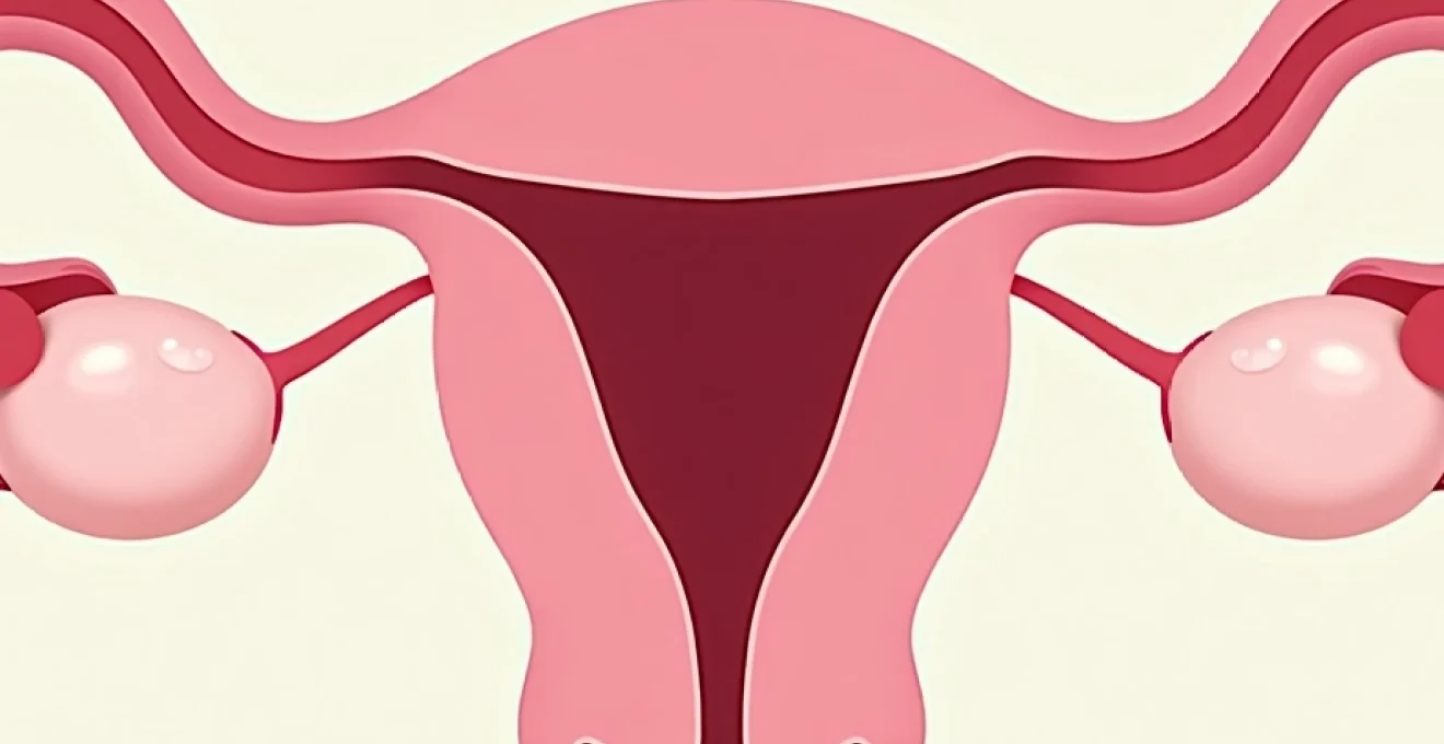
Discovering an ovarian cyst during a routine ultrasound examination can understandably cause concern for many women. The question of whether a 2.3 centimetre ovarian cyst represents a normal finding requires careful consideration of multiple clinical factors including patient age, menopausal status, cyst characteristics, and associated symptoms. In contemporary gynaecological practice, the management of small ovarian cysts has evolved significantly, with evidence-based guidelines providing clear frameworks for assessment and monitoring. Understanding the clinical significance of a 2.3cm ovarian cyst involves examining both the physiological processes that create these fluid-filled structures and the sophisticated diagnostic criteria used to distinguish benign lesions from those requiring surgical intervention.
Ovarian cyst classification and size parameters in reproductive medicine
The classification of ovarian cysts represents a fundamental cornerstone in gynaecological practice, with size parameters serving as crucial determinants for clinical management pathways. Modern reproductive medicine recognises two primary categories of ovarian cysts: functional cysts that arise from normal ovarian physiology and pathological cysts that develop through abnormal cellular processes. This distinction carries profound implications for patient counselling, monitoring protocols, and therapeutic decision-making.
Functional cyst categories: follicular and corpus luteum variations
Functional ovarian cysts emerge as natural consequences of the menstrual cycle, representing temporary enlargements of normal ovarian structures. Follicular cysts develop when dominant follicles fail to rupture during ovulation, continuing to grow beyond their typical 20-25mm diameter. These cysts typically measure between 2.5 and 8 centimetres, making a 2.3cm lesion slightly smaller than the conventional threshold for follicular cyst diagnosis.
Corpus luteum cysts form when the post-ovulatory corpus luteum fails to involute appropriately, accumulating fluid and potentially reaching sizes of 3-10 centimetres. The temporal relationship between cyst detection and menstrual cycle timing often provides valuable diagnostic clues, as functional cysts demonstrate characteristic patterns of growth and resolution correlating with hormonal fluctuations.
Research indicates that approximately 95% of ovarian cysts in premenopausal women represent functional variants, with the majority resolving spontaneously within 2-3 menstrual cycles.
Pathological cyst types: dermoid, endometrioma, and cystadenoma distinctions
Pathological ovarian cysts encompass a diverse spectrum of lesions arising from abnormal cellular proliferation or ectopic tissue deposition. Dermoid cysts (teratomas) contain mature tissues including hair, teeth, and sebaceous material, typically growing slowly at rates of 1-2mm annually. These lesions rarely present at 2.3cm diameter, as they usually achieve clinical recognition at larger sizes due to their distinctive ultrasonographic appearances.
Endometriomas develop from ectopic endometrial tissue implantation within ovarian parenchyma, creating characteristic “chocolate cysts” filled with altered blood products. While endometriomas can present at any size, lesions measuring 2.3cm may represent early-stage disease requiring careful monitoring for progression and associated symptoms.
Radiological measurement standards using transvaginal ultrasound protocols
Contemporary radiological assessment employs standardised transvaginal ultrasound protocols for precise ovarian cyst measurement and characterisation. The three-dimensional measurement approach utilises length, width, and anteroposterior dimensions to calculate mean diameter and volume parameters. A 2.3cm measurement typically represents the largest single dimension, though comprehensive assessment requires evaluation of all three planes for accurate volume determination.
High-frequency transvaginal probes provide superior resolution compared to transabdominal approaches, enabling detailed assessment of internal cyst architecture, wall thickness, and the presence of solid components or septations. These morphological features prove essential for risk stratification and management planning.
IOTA simple rules criteria for benign ovarian lesion assessment
The International Ovarian Tumour Analysis (IOTA) Simple Rules provide evidence-based criteria for distinguishing benign from malignant ovarian lesions. For benign classification, cysts must demonstrate unilocular appearance, absence of solid components, maximum diameter less than 10cm, absence of irregular thick septa, and minimal colour Doppler signal. A 2.3cm simple cyst meeting these criteria carries an extremely low malignancy risk, particularly in premenopausal women.
| IOTA Benign Features | IOTA Malignant Features |
|---|---|
| Unilocular cyst | Irregular solid tumour |
| Solid components <7mm | Ascites present |
| Smooth multilocular <10cm | ≥4 papillary structures |
| No blood flow | Irregular multilocular solid |
| Regular thin septa | Dense blood flow |
Clinical significance of 2.3 centimetre ovarian cysts in gynaecological practice
The clinical significance of a 2.3cm ovarian cyst varies considerably based on patient demographics, presenting symptoms, and associated risk factors. In routine gynaecological practice, such small cysts frequently represent incidental findings during pelvic imaging performed for unrelated indications. The challenge lies in determining which lesions require active monitoring versus those warranting immediate intervention or specialist referral.
Premenopausal versus postmenopausal risk stratification protocols
Age and menopausal status fundamentally influence risk assessment protocols for ovarian cysts. In premenopausal women, a 2.3cm simple cyst typically represents a physiological variant requiring minimal intervention. The ongoing ovarian activity creates numerous follicular structures, making small cysts extraordinarily common findings. Hormonal fluctuations throughout the menstrual cycle frequently cause these lesions to appear and disappear spontaneously.
Conversely, postmenopausal women face elevated malignancy risks even with smaller ovarian lesions. The absence of active folliculogenesis means any ovarian enlargement warrants careful evaluation. Current guidelines recommend that postmenopausal women with cysts exceeding 1cm undergo comprehensive assessment including tumour marker analysis and consideration for specialist referral.
Studies demonstrate that malignancy rates in postmenopausal ovarian cysts increase from 0.3% for lesions under 5cm to 35% for complex lesions exceeding 5cm diameter.
RCOG guidelines for small ovarian cyst management pathways
The Royal College of Obstetricians and Gynaecologists provides structured pathways for small ovarian cyst management, emphasising conservative approaches for appropriately selected patients. For premenopausal women with simple cysts measuring less than 5cm, including 2.3cm lesions, the recommended approach involves expectant management with follow-up ultrasound at 6-12 weeks to confirm resolution.
These guidelines acknowledge that functional cysts resolve spontaneously in approximately 70% of cases within two menstrual cycles. The decision to pursue active monitoring versus immediate intervention depends on symptom severity, patient anxiety levels, and individual risk factors including family history and genetic predisposition.
CA-125 tumour marker correlation with cyst size parameters
Serum CA-125 measurements provide valuable supplementary information for ovarian cyst assessment, though interpretation requires careful consideration of patient factors and clinical context. For 2.3cm cysts, elevated CA-125 levels may indicate underlying pathology, particularly in postmenopausal women where baseline values typically remain below 35 IU/mL.
However, numerous benign conditions can elevate CA-125 levels, including endometriosis, pelvic inflammatory disease, and even menstruation itself. In premenopausal women, CA-125 elevation alone does not justify surgical intervention for small cysts, but it may warrant enhanced monitoring protocols and multidisciplinary team discussion.
Risk of malignancy index (RMI) calculation for 2.3cm lesions
The Risk of Malignancy Index incorporates ultrasound findings, CA-125 levels, and menopausal status to generate numerical risk scores for ovarian lesions. For a 2.3cm simple cyst in a premenopausal woman with normal CA-125, the RMI score typically falls well below the 200 threshold indicating low malignancy risk. This quantitative approach provides objective data supporting conservative management strategies.
The RMI calculation proves particularly valuable for borderline cases where clinical features create diagnostic uncertainty. By combining multiple risk factors into a single numerical score, clinicians can make evidence-based decisions regarding the necessity for surgical exploration versus continued observation.
Diagnostic imaging protocols for small ovarian cysts
Contemporary diagnostic imaging protocols for small ovarian cysts emphasise multimodal approaches combining high-resolution ultrasound, advanced Doppler techniques, and selective use of cross-sectional imaging. The 2.3cm size threshold places these lesions within an intermediate category where basic ultrasound assessment typically provides sufficient information for clinical decision-making, though complex cases may require additional imaging modalities.
Transvaginal ultrasound doppler flow assessment techniques
Transvaginal ultrasound remains the gold standard initial imaging modality for ovarian cyst evaluation, offering superior resolution and detailed morphological assessment capabilities. Colour Doppler ultrasound provides crucial information regarding vascular patterns within cyst walls and solid components, helping distinguish benign from malignant lesions. For 2.3cm cysts, the absence of internal blood flow strongly supports benign diagnosis.
Power Doppler imaging demonstrates enhanced sensitivity for detecting low-velocity blood flow, particularly valuable when assessing small lesions where conventional colour Doppler may prove inadequate. The resistance index and pulsatility index measurements derived from spectral Doppler analysis provide quantitative vascular parameters that correlate with malignancy risk in larger studies.
MRI pelvis indications using T1 and T2-Weighted sequences
Magnetic resonance imaging serves as a problem-solving tool when ultrasound findings remain inconclusive or when cyst characteristics suggest specific pathological entities requiring tissue characterisation. For 2.3cm lesions, MRI indications typically include suspected endometriomas, dermoid cysts, or complex multilocular lesions requiring detailed internal architecture assessment.
T1-weighted sequences excel at detecting haemorrhage and fat content, while T2-weighted images provide superior soft tissue contrast for evaluating cyst contents and wall characteristics. The multiplanar imaging capability and superior tissue contrast resolution make MRI particularly valuable for distinguishing between different cyst types when ultrasound appearances remain ambiguous.
CT Abdomen-Pelvis contrast enhancement patterns
Computed tomography typically plays a limited role in routine 2.3cm ovarian cyst assessment, primarily reserved for cases where malignancy suspicion exists or when comprehensive abdominal evaluation becomes necessary. Contrast enhancement patterns within cyst walls or solid components can provide valuable information regarding vascularity and potential malignant transformation.
The superior ability of CT to detect peritoneal deposits, lymphadenopathy, and distant metastases makes it valuable for staging purposes when malignancy concerns arise. However, for small simple cysts in asymptomatic patients, CT imaging rarely provides additional clinically relevant information beyond high-quality ultrasound assessment.
Conservative management strategies for asymptomatic 2.3cm ovarian cysts
Conservative management approaches for asymptomatic 2.3cm ovarian cysts have gained widespread acceptance based on extensive research demonstrating excellent outcomes with expectant management strategies. The fundamental principle underlying conservative approaches recognises that the vast majority of small ovarian cysts represent benign entities that resolve spontaneously without intervention. This paradigm shift from aggressive surgical management to thoughtful observation reflects evolving understanding of ovarian cyst natural history and the potential risks associated with unnecessary surgical procedures.
The conservative management protocol typically begins with comprehensive patient counselling regarding cyst characteristics, expected outcomes, and warning signs requiring immediate medical attention. Patient education plays a crucial role in successful conservative management, as informed patients demonstrate better compliance with follow-up schedules and appropriate recognition of concerning symptoms. The psychological aspects of cyst diagnosis require careful attention, as many patients experience significant anxiety despite receiving reassurance about benign findings.
Follow-up imaging schedules vary depending on patient factors and cyst characteristics, though most protocols recommend initial repeat ultrasound at 6-8 weeks for premenopausal women with simple cysts. The timing allows for at least one complete menstrual cycle, providing opportunity for functional cysts to resolve naturally. If the cyst persists but remains unchanged in size and appearance, extended monitoring intervals of 3-6 months may prove appropriate, particularly when patients remain asymptomatic.
Hormonal contraception represents an important consideration in conservative management strategies, though its role remains somewhat controversial. While combined oral contraceptives do not accelerate existing cyst resolution, they may prevent formation of new functional cysts by suppressing ovulation. This approach proves particularly beneficial for women with recurrent functional cysts or those requiring long-term monitoring of persistent lesions.
Long-term follow-up studies indicate that 85-90% of simple ovarian cysts measuring less than 3cm resolve spontaneously within 6 months, supporting conservative management as the preferred initial approach.
Pain management during conservative monitoring typically involves simple analgesics and lifestyle modifications. Patients should receive clear instructions about activity restrictions, particularly avoiding activities that might increase intra-abdominal pressure or risk cyst rupture. However, most daily activities can continue normally, and excessive restrictions may unnecessarily impact quality of life.
Surgical intervention thresholds: laparoscopic cystectomy considerations
Surgical intervention for 2.3cm ovarian cysts remains relatively uncommon, reserved primarily for cases where conservative management fails or specific clinical circumstances mandate tissue diagnosis. The decision-making process for surgical intervention requires careful weighing of potential benefits against inherent operative risks, particularly considering that most small cysts prove benign upon histological examination. Laparoscopic cystectomy has emerged as the preferred surgical approach when intervention becomes necessary, offering advantages of minimal invasive access, reduced post-operative morbidity, and preservation of healthy ovarian tissue.
The primary indications for surgical intervention in 2.3cm cysts include persistence beyond 3-6 months despite appropriate monitoring, development of concerning ultrasonographic features such as solid components or thick septations, and onset of significant symptoms including persistent pelvic pain or pressure sensations. Patient factors such as strong family history of ovarian cancer, presence of BRCA gene mutations, or extreme patient anxiety despite appropriate counselling may also influence surgical decision-making.
Laparoscopic surgical techniques for small cysts emphasise ovarian preservation through careful tissue handling and precise dissection techniques. The stripping technique involves identifying the correct tissue plane between cyst wall and normal ovarian parenchyma, allowing complete cyst removal while minimising damage to surrounding healthy tissue. Experienced laparoscopic surgeons can typically remove 2.3cm cysts with minimal loss of ovarian reserve, though pre-operative counselling should address potential risks including inadvertent removal of healthy ovarian tissue.
Post-operative outcomes following laparoscopic cystectomy for small ovarian cysts generally demonstrate excellent results with low complication rates and rapid recovery times. Most patients experience resolution of pre-operative symptoms when present, and histological examination provides definitive tissue diagnosis. The procedure typically requires only day-case admission, allowing patients to return to normal activities within 1-2 weeks. However, the potential impact on ovarian reserve remains a consideration, particularly for women planning future pregnancies or those with already diminished ovarian function.
Alternative surgical approaches such as cyst aspiration or sclerotherapy have been investigated for small benign cysts, though these techniques carry higher recurrence rates and lack the diagnostic advantages of complete surgical excision. The inability to obtain tissue for histological examination represents a significant limitation of aspiration techniques, particularly given the occasional unexpected finding of malignancy even in apparently benign small lesions. Current evidence strongly supports la
paroscopic cystectomy as the gold standard surgical approach when intervention becomes necessary for small ovarian cysts.
The timing of surgical intervention requires individualised assessment considering patient age, fertility goals, and symptom severity. For women actively trying to conceive, surgical delays may prove appropriate if conservative management shows signs of success, as even minor surgical trauma to ovarian tissue can potentially impact fertility outcomes. Conversely, women who have completed their families or those experiencing significant quality of life impairment may benefit from earlier surgical intervention to achieve symptom resolution and provide peace of mind through definitive histological diagnosis.
Intraoperative findings during laparoscopic cystectomy for 2.3cm lesions frequently reveal straightforward surgical anatomy with clear tissue planes facilitating complete cyst removal. The small size typically allows for precise dissection without requiring complex reconstructive techniques. Frozen section analysis during surgery provides immediate histological feedback when malignancy concerns exist, though this investigation proves necessary in fewer than 5% of cases involving small simple cysts in premenopausal women.
Post-operative monitoring following surgical intervention involves routine follow-up ultrasound at 6-12 weeks to confirm complete cyst removal and assess ovarian healing. Long-term surveillance protocols depend on final histological results, with benign findings typically requiring no further specific monitoring beyond routine gynaecological care. The excellent outcomes associated with laparoscopic management of small ovarian cysts have established this approach as a safe and effective option when conservative management proves insufficient or clinical circumstances mandate surgical exploration.
Meta-analyses of laparoscopic cystectomy outcomes demonstrate 95% complete cyst removal rates with less than 2% major complication rates, supporting surgical intervention when clinically indicated for small ovarian lesions.
The integration of conservative and surgical management strategies creates a comprehensive approach to 2.3cm ovarian cyst management that prioritises patient safety while minimising unnecessary interventions. This balanced approach recognises that while most small cysts prove benign and self-limiting, appropriate surgical intervention remains essential for selected cases where conservative management fails to achieve desired outcomes or clinical factors suggest potential malignancy risk.