Spinal hemangiomas represent the most prevalent benign vascular tumours affecting the vertebral column, occurring in approximately 10% of the global population. While these abnormal blood vessel growths typically remain asymptomatic throughout their course, a small subset develops aggressive characteristics that can produce significant neurological complications. Understanding the complex interplay between hemangioma pathophysiology and neural compression mechanisms becomes crucial when these seemingly innocuous lesions begin to compromise spinal cord function or nerve root integrity.
The transition from an incidental radiological finding to a clinically significant neurological condition often catches both patients and healthcare providers off guard. Less than 5% of spinal hemangiomas ever produce symptoms, yet when they do, the presentations can range from mild radicular pain to devastating myelopathy with paralysis. Recognition of early neurological warning signs enables timely intervention and prevents irreversible spinal cord damage that can profoundly impact quality of life.
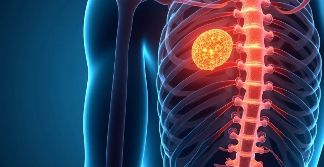
Pathophysiology and anatomical distribution of spinal hemangiomas
The development of neurologically symptomatic spinal hemangiomas involves a complex cascade of vascular proliferation, osseous expansion, and subsequent neural compression. These benign tumours originate from abnormal proliferation of endothelial cells within the vertebral body, creating a network of dilated vascular channels that gradually expand beyond their normal anatomical boundaries. The progressive enlargement of these vascular spaces weakens the structural integrity of the vertebral bone, leading to characteristic radiographic appearances and, in aggressive cases, epidural extension.
Vertebral body hemangioma vascular architecture and neural compression mechanisms
The intricate vascular architecture of symptomatic hemangiomas demonstrates a honeycomb or lattice-like pattern on imaging studies, reflecting the underlying pathological organisation of dilated sinusoidal spaces. These vascular channels can expand rapidly, particularly during periods of increased blood flow or hormonal fluctuation, creating a mass effect that extends beyond the confines of the vertebral body. The mechanical compression of neural structures occurs through multiple mechanisms, including direct pressure from the expanding tumour mass, secondary spinal stenosis, and compromise of the neural foramen.
Thoracic spine T6-T12 predilection sites and associated neurological risk factors
Thoracic vertebrae between T6 and T12 demonstrate the highest propensity for harboring clinically significant hemangiomas, with this region accounting for approximately 60% of all symptomatic cases. The relatively narrow spinal canal diameter in the thoracic region, combined with the critical vascular supply to the thoracic spinal cord, creates a perfect storm for neurological compromise. Age-related changes in bone density and hormonal influences, particularly oestrogen fluctuations in postmenopausal women, can accelerate hemangioma growth and increase the likelihood of symptom development.
Epidural extension patterns and spinal canal stenosis development
When hemangiomas breach the posterior cortex of the vertebral body, epidural extension represents the most concerning complication from a neurological standpoint. This extraosseous growth pattern typically manifests as a soft tissue mass that directly compresses the thecal sac, leading to varying degrees of spinal canal stenosis. The rate of epidural extension can be unpredictable, with some cases demonstrating rapid progression over weeks to months, whilst others maintain stable configurations for years before suddenly expanding.
Osseous expansion effects on adjacent nerve root foramina
The progressive expansion of hemangiomatous tissue within the vertebral body inevitably affects the dimensions of adjacent neural foramina, creating compression syndromes that manifest as radicular symptoms. This foraminal stenosis develops through both direct tumour extension and secondary inflammatory responses that further narrow these critical anatomical spaces. The resulting nerve root compression produces characteristic dermatomal pain patterns and motor weakness that can be mistaken for more common conditions such as disc herniation or degenerative spinal disease.
Clinical manifestations of compressive spinal hemangioma symptoms
The neurological presentation of symptomatic spinal hemangiomas encompasses a broad spectrum of clinical findings that reflect the anatomical level of compression and the extent of neural involvement. Early recognition of these symptoms becomes paramount, as prompt intervention can prevent irreversible neurological damage and preserve functional capacity. The insidious onset of symptoms often delays diagnosis, as patients and clinicians may initially attribute complaints to more common spinal conditions or age-related degenerative changes.
Progressive myelopathy secondary to cord compression syndromes
Myelopathy represents the most serious neurological complication of spinal hemangiomas, developing when tumour expansion or epidural extension creates significant spinal cord compression. The clinical presentation typically follows a predictable pattern, beginning with subtle gait disturbances and progressing to more obvious motor and sensory deficits. Patients may initially notice difficulty with fine motor tasks, such as buttoning clothes or writing, before developing more pronounced weakness and coordination problems. The hallmark signs of myelopathy include hyperreflexia, pathological reflexes such as Babinski signs, and spasticity that predominantly affects the lower extremities.
Radicular pain distribution following dermatome patterns
Radicular symptoms arising from hemangioma-related nerve root compression typically manifest as sharp, shooting pain that follows specific dermatomal distributions. This pain often demonstrates characteristic features that distinguish it from mechanical back pain, including exacerbation with coughing, sneezing, or Valsalva manoeuvres. The intensity can vary significantly throughout the day, with many patients reporting increased discomfort during periods of increased blood flow or when assuming certain positions that enhance venous congestion within the hemangioma.
Motor weakness presentations in upper and lower extremities
Motor weakness patterns in hemangioma-related neurological compression depend heavily on the anatomical level of involvement and the specific neural structures affected. Cervical hemangiomas may produce upper extremity weakness that follows myotomal patterns, whilst thoracic lesions typically manifest as lower extremity motor deficits. The progression of motor weakness can be particularly rapid in cases with aggressive hemangioma growth, sometimes advancing from subtle weakness to complete paralysis within weeks if left untreated.
The rapid progression from mild symptoms to severe neurological deficits in aggressive spinal hemangiomas underscores the critical importance of early recognition and prompt intervention to preserve neural function.
Sensory disturbances and paraesthesia localisation patterns
Sensory symptoms associated with spinal hemangioma compression typically begin as subtle tingling or numbness that gradually progresses to more pronounced sensory loss. These disturbances often follow anatomical patterns that correspond to the level of compression, with patients describing burning sensations, pins and needles, or complete numbness in affected dermatomes. The sensory level can serve as a valuable diagnostic clue, particularly when it corresponds to the anatomical location of the hemangioma on imaging studies.
Bowel and bladder dysfunction in cauda equina involvement
Involvement of the cauda equina by lumbar hemangiomas can produce devastating bowel and bladder dysfunction that represents a true neurological emergency. These symptoms typically develop gradually, beginning with subtle changes in urinary hesitancy or bowel habits before progressing to complete incontinence or retention. Cauda equina syndrome requires immediate surgical decompression to prevent permanent neurological disability and preserve sphincter function.
Differential diagnosis considerations for spinal hemangioma neurological presentations
The neurological symptoms produced by spinal hemangiomas can closely mimic numerous other spinal pathologies, creating diagnostic challenges that may delay appropriate treatment. Clinicians must maintain a high index of suspicion for hemangioma-related compression, particularly in middle-aged women with progressive neurological symptoms and characteristic imaging findings. The differential diagnosis encompasses both benign and malignant conditions that can produce similar clinical presentations through various mechanisms of neural compression.
Herniated intervertebral discs represent the most common alternative diagnosis, particularly when patients present with radicular pain patterns that follow dermatomal distributions. However, the age demographics and imaging characteristics often help distinguish between these conditions, as hemangiomas typically affect older individuals and demonstrate characteristic vascular patterns on magnetic resonance imaging. Spinal metastases pose another significant diagnostic consideration, especially in patients with known malignancies or constitutional symptoms suggestive of systemic disease.
Degenerative spinal stenosis frequently produces similar neurological symptoms, particularly in the elderly population where both conditions may coexist. The key differentiating features include the characteristic honeycomb appearance of hemangiomas on imaging studies and the absence of the typical claudication symptoms seen with degenerative stenosis. Multiple sclerosis and other demyelinating conditions can occasionally present with similar myelopathic symptoms, requiring careful evaluation of imaging studies and cerebrospinal fluid analysis to establish the correct diagnosis.
Advanced neuroimaging assessment protocols for symptomatic hemangiomas
Comprehensive neuroimaging evaluation forms the cornerstone of diagnosis and treatment planning for neurologically symptomatic spinal hemangiomas. The multimodal imaging approach combines various techniques to provide detailed anatomical information about tumour characteristics, extent of neural compression, and vascular anatomy. Modern imaging protocols have evolved to address the specific challenges posed by these vascular lesions, incorporating both static and dynamic studies to fully characterise their impact on neural structures.
MRI T1 and T2 weighted sequence interpretation for neural compression
Magnetic resonance imaging serves as the gold standard for evaluating spinal hemangiomas and their neurological impact, with specific pulse sequences providing complementary information about tumour characteristics and neural compression. T1-weighted sequences typically demonstrate the characteristic high signal intensity of hemangiomas due to their fat content, creating the pathognomonic “polka-dot” appearance on axial images. T2-weighted sequences reveal the extent of epidural extension and the degree of spinal cord compression, often showing high signal changes within the spinal cord itself in cases of significant compression. The combination of T1 and T2 findings allows for accurate assessment of both tumour morphology and its impact on surrounding neural structures.
CT myelography applications in epidural extension evaluation
Computed tomography myelography provides superior detail of epidural extension patterns and the degree of thecal sac compression, particularly in cases where MRI findings are equivocal or contraindicated. This invasive imaging technique involves intrathecal contrast injection followed by CT imaging, creating detailed images of the spinal canal and nerve root sleeves. The technique proves particularly valuable in surgical planning, as it clearly delineates the relationship between the hemangioma and critical neural structures that must be preserved during decompressive procedures.
Dynamic MRI studies for Postural-Dependent symptom assessment
Dynamic or flexion-extension MRI studies can reveal position-dependent changes in spinal canal dimensions and neural compression that may not be apparent on standard supine imaging. These studies prove particularly valuable in patients whose symptoms demonstrate positional variation, as they can identify dynamic stenosis caused by hemangioma expansion during specific spinal movements. The ability to correlate symptoms with imaging findings in various spinal positions enhances diagnostic accuracy and treatment planning precision.
Spinal angiography indications for Pre-Surgical planning
Spinal angiography becomes essential in cases requiring surgical intervention or embolisation therapy, as it provides detailed information about the vascular architecture and feeding vessels of the hemangioma. This invasive procedure identifies the arterial supply and venous drainage patterns, which are crucial for planning safe surgical approaches and embolisation strategies. The study also helps identify potential anastomoses with critical spinal cord vessels, preventing inadvertent injury to the anterior spinal artery or other vital vascular structures during treatment.
Advanced neuroimaging protocols must be tailored to each individual case, considering the specific clinical presentation, anatomical location, and proposed treatment strategy to optimise diagnostic accuracy and therapeutic outcomes.
Treatment algorithms for neurologically symptomatic spinal hemangiomas
The management of neurologically symptomatic spinal hemangiomas requires a multidisciplinary approach that considers the severity of symptoms, rate of progression, anatomical location, and patient factors when selecting appropriate treatment modalities. Treatment algorithms have evolved significantly over the past decade, incorporating minimally invasive techniques alongside traditional surgical approaches to optimise patient outcomes whilst minimising procedural risks. The urgency of intervention depends heavily on the degree of neurological compromise, with acute presentations requiring emergency treatment to prevent irreversible neural damage.
Percutaneous vertebroplasty techniques and neurological outcome metrics
Percutaneous vertebroplasty has emerged as a first-line treatment option for many symptomatic spinal hemangiomas, particularly those causing pain without significant neural compression. This minimally invasive procedure involves the injection of polymethylmethacrylate cement directly into the hemangiomatous vertebral body, stabilising the bone and reducing pain through thermal and mechanical effects. The neurological outcomes following vertebroplasty demonstrate significant improvement in pain scores and functional capacity, with most patients experiencing symptom relief within 24-48 hours of the procedure. Success rates exceed 85% for appropriately selected cases, with minimal complications when performed by experienced interventionalists.
The technique requires careful fluoroscopic guidance to ensure accurate cement placement whilst avoiding extravasation into the spinal canal or neural foramina. Pre-procedural planning involves detailed review of cross-sectional imaging to identify safe trajectories and appropriate cement volumes. Patient selection criteria include the absence of significant epidural extension, adequate bone corridors for needle placement, and realistic expectations regarding symptom improvement. Long-term follow-up studies demonstrate sustained pain relief and functional improvement in the majority of patients, with low rates of adjacent level problems or cement-related complications.
Surgical decompression approaches via laminectomy and corpectomy
Surgical intervention becomes necessary in cases with significant neural compression, progressive neurological deficits, or failure of conservative treatment modalities. The choice between laminectomy and corpectomy depends on the location and extent of neural compression, with posterior approaches preferred for primarily epidural disease and anterior or combined approaches reserved for cases with significant vertebral body involvement. Laminectomy with tumour debulking provides excellent exposure for epidural extension removal whilst preserving spinal stability in most cases.
Corpectomy procedures, whilst more technically demanding, offer complete tumour removal and immediate spinal cord decompression in cases with extensive vertebral body involvement. These procedures typically require anterior approach with reconstruction using titanium cages or bone grafts, followed by posterior instrumentation for stability. The neurological recovery rates following surgical decompression depend heavily on the preoperative neurological status, with better outcomes achieved when intervention occurs before irreversible neural damage develops.
Radiation therapy protocols for inoperable lesions
Radiation therapy serves as an alternative treatment modality for patients who are poor surgical candidates or have lesions in locations that preclude safe surgical intervention. Modern radiation techniques, including intensity-modulated radiation therapy (IMRT) and stereotactic body radiation therapy (SBRT), allow for precise dose delivery whilst minimising exposure to critical structures. Treatment protocols typically involve fractionated doses totalling 20-30 Gy delivered over multiple sessions to optimise tumour response whilst reducing radiation-related complications.
The mechanism of action involves endothelial cell damage and subsequent vascular thrombosis, leading to gradual tumour regression and symptom improvement over weeks to months. Response rates to radiation therapy approach 70-80% for pain relief, though neurological recovery may be more limited compared to surgical intervention. Long-term follow-up is essential to monitor for delayed radiation effects and ensure sustained tumour control.
Embolisation procedures using polyvinyl alcohol particles
Transcatheter embolisation represents a sophisticated interventional technique that targets the vascular supply of spinal hemangiomas, inducing thrombosis and subsequent tumour regression. The procedure involves selective catheterisation of feeding arteries followed by deployment of polyvinyl alcohol particles, coils, or liquid embolic agents to occlude the tumour’s blood supply. Pre-procedural spinal angiography identifies the vascular anatomy and ensures that critical spinal cord vessels are not inadvertently compromised during embolisation.
Success rates for embolisation procedures vary depending on the tumour’s vascular architecture and the completeness of devascularisation achieved. The combination of embolisation with subsequent surgical intervention or vertebr
oplasty can significantly enhance the safety and efficacy of both procedures, reducing intraoperative bleeding and improving visualisation during surgical decompression.The technical aspects of embolisation require careful consideration of particle size and embolic agent selection to achieve optimal tumour devascularisation whilst avoiding non-target embolisation. Polyvinyl alcohol particles ranging from 100-300 micrometers provide excellent penetration into the tumour neovasculature whilst minimising the risk of passing through arteriovenous shunts. Post-embolisation syndrome represents a common but manageable complication, characterised by fever, pain, and nausea that typically resolves within 48-72 hours with supportive care.
The multidisciplinary approach to treating neurologically symptomatic spinal hemangiomas requires careful coordination between interventional radiologists, neurosurgeons, and radiation oncologists to optimise patient outcomes. Treatment selection algorithms must consider multiple factors including patient age, comorbidities, symptom severity, rate of progression, and anatomical characteristics of the lesion. Emergency presentations with acute neurological deterioration may require immediate surgical decompression, whilst more stable cases can undergo staged treatment with embolisation followed by definitive therapy.
Long-term monitoring protocols following treatment involve regular neurological assessments and imaging studies to evaluate treatment response and detect potential complications. Most patients experience significant symptom improvement within the first few weeks following treatment, though complete neurological recovery may take several months depending on the degree of preoperative neural compromise. Recurrence rates remain low with appropriate treatment selection, though long-term surveillance remains essential to detect delayed complications or treatment failure.
The evolution of treatment algorithms for symptomatic spinal hemangiomas reflects the integration of minimally invasive techniques with traditional surgical approaches, enabling personalised treatment strategies that optimise outcomes whilst minimising procedural risks.
Quality of life assessments following treatment demonstrate significant improvements in pain scores, functional capacity, and patient satisfaction across all treatment modalities. The choice of treatment approach should be individualised based on patient-specific factors, institutional expertise, and the urgency of neurological symptoms. Shared decision-making between patients and their healthcare teams ensures that treatment selection aligns with individual goals and expectations whilst achieving optimal neurological outcomes.