Vulvar fissures and labial tears present a distressing yet surprisingly common gynaecological concern that affects women across all age groups. These delicate tissue injuries, often described as feeling like paper cuts on the sensitive vulvar area, can cause significant discomfort during daily activities, urination, and intimate moments. The characteristic sharp, stinging sensation these lesions produce belies their typically small size, creating disproportionate pain that can substantially impact quality of life. Understanding the multifaceted nature of these injuries is crucial for both healthcare providers and patients, as the underlying causes range from simple mechanical trauma to complex autoimmune conditions requiring specialised treatment approaches.
Vulvar fissures and mucosal tears: clinical presentation and anatomical distribution
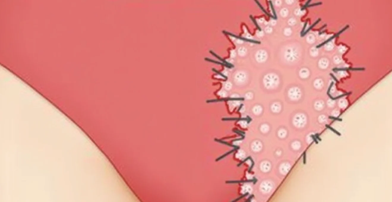
Vulvar fissures manifest as linear splits or cracks in the delicate skin and mucous membranes surrounding the external female genitalia. These lesions typically measure between 2-10 millimetres in length and can vary significantly in depth, from superficial epithelial breaks to deeper wounds extending into the submucosal layers. The clinical presentation often includes acute onset pain, particularly during activities that stretch the affected tissue, accompanied by occasional light bleeding or spotting.
The anatomical distribution of these fissures follows predictable patterns based on areas of greatest mechanical stress and tissue vulnerability. Research indicates that approximately 60-70% of vulvar fissures occur in the posterior vestibule and perineal area, where thin mucosal tissues are subjected to the greatest stretching forces during sexual activity or childbirth. The remaining cases distribute across the labial folds, clitoral hood, and anterior vestibule, with each location presenting distinct challenges for diagnosis and management.
Labial majora fissuring patterns and depth assessment
Fissures affecting the labial majora typically occur along the natural fold lines where keratinised skin meets the more delicate mucosal surfaces. These lesions often present as bilateral symmetrical tears, particularly when associated with systemic conditions such as lichen sclerosus or hormonal deficiency states. The depth assessment requires careful visual examination, as superficial tears affecting only the stratum corneum heal rapidly with conservative management, whilst deeper fissures extending into the dermis may require more aggressive intervention.
Vestibular mucosa laceration characteristics
The vestibular mucosa, comprising the area between the labia minora and the vaginal entrance, represents the most vulnerable site for fissure development. These tears characteristically appear as linear erosions with well-defined borders, often accompanied by localised erythema and oedema. The vestibular tissue’s rich nerve supply explains the intense pain associated with even minor fissures in this region, creating symptoms that seem disproportionate to the visible injury.
Posterior fourchette split distribution in Reproductive-Age women
Posterior fourchette fissures occur predominantly in sexually active women of reproductive age, with studies showing peak incidence between ages 25-35 years. These splits typically measure 5-8 millimetres in length and occur at the midline commissure where the labia minora meet posteriorly. The tissue architecture in this region, designed for elasticity during sexual activity, can become compromised by repeated trauma or underlying inflammatory conditions, leading to recurrent splitting patterns.
Clitoral hood microtrauma and associated bleeding patterns
Microtrauma to the clitoral hood presents unique diagnostic challenges due to the area’s complex anatomy and varying degrees of coverage. These injuries often result from aggressive manipulation during sexual activity or improper personal hygiene practices. The associated bleeding patterns typically manifest as pinpoint haemorrhages or light spotting, distinguishing them from more serious vascular injuries requiring immediate medical attention.
Infectious aetiologies: bacterial, viral, and fungal pathogens
Infectious agents represent a significant category of causative factors in vulvar fissuring, with various pathogens creating tissue inflammation and subsequent breakdown. The compromised barrier function resulting from these infections predisposes the vulvar tissues to mechanical trauma and delayed healing. Understanding the specific pathogenic mechanisms helps clinicians develop targeted therapeutic approaches and prevent recurrent episodes.
Studies demonstrate that approximately 40% of recurrent vulvar fissures have an underlying infectious component, with Candida species accounting for nearly half of these cases.
Herpes simplex virus type 1 and type 2 ulcerative lesions
Herpes simplex virus infections create characteristic vesicular lesions that rupture to form shallow, painful ulcers resembling fissures. HSV-1 and HSV-2 both demonstrate neurotropic properties, establishing latency in regional nerve ganglia and causing recurrent outbreaks. The initial presentation often includes prodromal symptoms such as tingling or burning sensations, followed by vesicle formation and subsequent ulceration. These viral lesions typically heal within 7-10 days but may leave areas of tissue weakness susceptible to future mechanical trauma.
Candida Albicans-Induced vulvar fissuring and pruritic response
Candida albicans infections create an inflammatory environment that compromises epithelial integrity and increases susceptibility to fissure formation. The characteristic intense pruritus associated with candidiasis leads to scratching behaviour that further traumatises already vulnerable tissues. Chronic candidiasis can result in persistent inflammation and tissue remodelling, creating areas of increased fragility prone to recurrent splitting. The cyclical nature of these infections, often correlating with menstrual cycles or antibiotic use, contributes to the chronic, recurrent pattern seen in many patients.
Group B streptococcus secondary bacterial infections
Group B Streptococcus (GBS) can establish secondary infections in pre-existing vulvar fissures, complicating the healing process and potentially leading to deeper tissue involvement. These gram-positive bacteria demonstrate particular affinity for compromised epithelial barriers, establishing biofilm formation that resists conventional antimicrobial therapy. The inflammatory response to GBS infection can result in tissue oedema and delayed wound closure, perpetuating the cycle of trauma and infection.
Human papillomavirus subtypes 6 and 11 tissue disruption
HPV subtypes 6 and 11, primarily associated with benign genital warts, can cause tissue architecture disruption that predisposes to fissure formation. The viral infection induces hyperkeratosis and cellular proliferation, creating areas of tissue irregularity and increased mechanical stress concentration. While these low-risk HPV types rarely progress to malignancy, their impact on tissue integrity can create chronic wounds that heal poorly without appropriate antiviral intervention.
Hormonal fluctuations and Oestrogen-Deficiency states
Hormonal influences play a pivotal role in maintaining vulvar tissue integrity and healing capacity. Oestrogen deficiency, whether physiological or iatrogenic, leads to significant changes in tissue architecture and mechanical properties. The vulvovaginal tissues contain abundant oestrogen receptors, and declining hormone levels result in decreased collagen synthesis, reduced tissue thickness, and compromised vascular supply. These changes create an environment where minor mechanical stresses can result in tissue breakdown and poor healing responses.
The temporal relationship between hormonal fluctuations and fissure development provides important diagnostic clues. Many women report symptom exacerbation during specific phases of their menstrual cycle, particularly the luteal phase when oestrogen levels decline. Similarly, life transitions associated with hormonal changes, such as perimenopause, postpartum periods, and certain contraceptive methods, correlate with increased fissure incidence and severity.
Postmenopausal vulvovaginal atrophy and tissue fragility
Postmenopausal vulvovaginal atrophy represents the most severe form of oestrogen-deficiency-related tissue changes. The lack of circulating oestrogen leads to progressive thinning of the epithelium, loss of glycogen stores, and decreased lactobacilli colonisation. These changes result in elevated vaginal pH, increased susceptibility to infection, and significantly reduced tissue elasticity. The combination of mechanical fragility and compromised immune function creates optimal conditions for recurrent fissure formation that responds poorly to conservative measures alone.
Lactational Amenorrhoea-Associated mucosal thinning
Breastfeeding women experience prolonged hypoestrogenic states due to prolactin-mediated suppression of gonadotropin-releasing hormone. This physiological adaptation ensures adequate milk production but compromises vulvovaginal tissue integrity. The mucosal thinning associated with lactational amenorrhoea typically resolves gradually following weaning, though some women may require topical oestrogen therapy to accelerate tissue recovery and prevent recurrent trauma.
Perimenarchal hormonal imbalances and vulvar sensitivity
Young women experiencing menarche and early menstrual cycles may encounter hormonal fluctuations that affect tissue integrity. The immature hypothalamic-pituitary-ovarian axis creates irregular hormone patterns that can result in periods of relative oestrogen deficiency. Additionally, the psychosocial stress associated with puberty and early sexual experiences may exacerbate tissue sensitivity and delay healing responses.
Contraceptive-induced oestradiol suppression effects
Modern low-dose combined oral contraceptives and progestin-only methods can significantly suppress endogenous oestradiol production. Some women demonstrate particular sensitivity to these hormonal changes, developing vulvar symptoms despite normal systemic hormone levels. Depot medroxyprogesterone acetate injections represent the most significant risk factor among contraceptive methods, with some studies reporting vulvar complaints in up to 15% of users within the first year of administration.
Dermatological conditions: lichen sclerosus and autoimmune vulvopathies
Dermatological conditions affecting the vulvar region create chronic inflammatory states that predispose to recurrent fissuring and poor wound healing. These conditions often present diagnostic challenges due to their subtle initial presentations and overlap with other vulvar disorders. Lichen sclerosus stands as the most significant dermatological cause of chronic vulvar fissuring, affecting approximately 1 in 900 women and demonstrating a bimodal age distribution with peaks in prepubertal girls and postmenopausal women.
The pathophysiology of autoimmune vulvopathies involves complex interactions between genetic predisposition, environmental triggers, and immune system dysfunction. These conditions create progressive tissue architecture changes, including epithelial thinning, dermal sclerosis, and compromised vascular supply. The resulting tissue fragility makes even minor mechanical stresses capable of causing significant trauma and delayed healing.
Early recognition and treatment of vulvar dermatological conditions can prevent irreversible architectural changes and reduce long-term morbidity associated with chronic fissuring.
Lichen sclerosus demonstrates characteristic histological features including hyperkeratosis, epithelial atrophy, and homogenisation of dermal collagen. The condition typically begins as small, white papules that coalesce into larger plaques with a distinctive “cigarette paper” appearance. The progressive nature of untreated lichen sclerosus can result in architectural distortion, including labial fusion and introital stenosis, creating mechanical factors that perpetuate the cycle of trauma and poor healing.
Other dermatological conditions contributing to vulvar fissuring include lichen planus, which creates erosive lesions and lacy white striations, and psoriasis, which can present atypically in the genital region without the characteristic silvery scales seen elsewhere. Contact dermatitis, whether allergic or irritant in nature, creates acute inflammatory responses that compromise epithelial barrier function and increase susceptibility to mechanical trauma. The diagnosis of these conditions often requires specialised testing, including patch testing for contact allergens and sometimes vulvar biopsy for definitive histological confirmation.
Mechanical trauma and sexual Activity-Related injuries
Mechanical trauma represents the most common immediate cause of vulvar fissures, particularly in sexually active women. The delicate balance between tissue elasticity and mechanical stress can be disrupted by various factors, including inadequate lubrication, aggressive sexual techniques, or anatomical variations that concentrate stress in vulnerable areas. Understanding the biomechanics of vulvar trauma helps clinicians provide targeted prevention strategies and identify patients at higher risk for recurrent injuries.
Sexual activity-related injuries demonstrate specific patterns based on the type and intensity of contact involved. Penetrative activities create the highest risk for posterior fourchette injuries, while external stimulation may result in clitoral hood or labial trauma. The use of sex toys, particularly those with rough textures or inappropriate sizes, can create localised areas of high mechanical stress leading to tissue breakdown. Insufficient arousal time compounds these risks by limiting natural lubrication and reducing tissue pliability.
Risk factors for mechanical trauma extend beyond sexual practices to include personal hygiene habits, clothing choices, and exercise activities. Aggressive cleansing with harsh soaps or abrasive materials can compromise the natural protective barriers of vulvar skin. Tight-fitting synthetic undergarments create chronic friction and moisture retention, establishing conditions favourable for tissue breakdown and delayed healing. High-impact exercise activities, particularly cycling and horseback riding, can create repetitive trauma that accumulates over time.
The healing response to mechanical trauma varies significantly based on individual factors including age, hormonal status, and overall health. Younger women with adequate oestrogen levels typically demonstrate rapid healing within 5-7 days, while postmenopausal women or those with compromised immune function may require weeks for complete resolution. The presence of concurrent infections or inflammatory conditions can significantly prolong healing times and increase the risk of recurrent trauma in the same location.
Differential diagnosis: bartholin’s cyst rupture and hidradenitis suppurativa
Accurate differential diagnosis of vulvar fissures requires careful consideration of various conditions that can present with similar symptoms of pain, discharge, and tissue breakdown. Bartholin’s cyst rupture can create sudden-onset vulvar pain and discharge that may be confused with infectious fissures, particularly when the rupture occurs through the skin surface rather than into the vaginal canal. The anatomical location of Bartholin’s glands at the posterolateral aspects of the vaginal introitus helps distinguish these lesions from typical fissure locations.
Hidradenitis suppurativa represents a chronic inflammatory condition affecting apocrine gland-bearing areas, including the vulva and groin. This condition creates recurrent painful nodules and sinus tracts that can rupture and drain, producing symptoms similar to infected vulvar fissures. The characteristic “double-ended” comedones and rope-like scarring patterns help distinguish hidradenitis suppurativa from simple fissures, though early stages of the disease may present diagnostic challenges.
Vulvar Behçet’s disease, though rare, can create aphthous-like ulcers that may be mistaken for severe fissures, particularly in patients with concurrent oral lesions or systemic symptoms.
Other conditions requiring consideration in the differential diagnosis include vulvar vestibulitis syndrome, which creates burning pain without visible fissures, and various malignancies that can present as non-healing ulcerative lesions. Crohn’s disease can create characteristic “knife-cut” fissures with undermined edges, typically occurring in patients with known inflammatory bowel disease or other systemic symptoms. The timing, location, and associated symptoms provide crucial diagnostic clues that guide appropriate investigation and treatment strategies.
Advanced diagnostic techniques may be necessary in challenging cases, including vulvoscopy for detailed visual assessment, microbiology sampling for infectious agents, and histopathological examination when malignancy or unusual inflammatory conditions are suspected. The integration of clinical findings with appropriate investigations ensures accurate diagnosis and optimal treatment outcomes for patients presenting with vulvar fissuring complaints.