Discovering unusual formations in stool that resemble branched roots or plant-like structures can be deeply concerning for anyone experiencing this phenomenon. These mysterious appearances, often described as having the texture of branched sticks or root systems, frequently prompt urgent questions about their origin and potential health implications. The human digestive system processes a remarkable variety of materials daily, and the appearance of unfamiliar objects in faecal matter can indicate several distinct possibilities ranging from completely benign dietary residues to parasitic infections requiring medical attention.
Understanding what creates these root-like appearances requires examining multiple factors including recent dietary intake, digestive health status, and potential exposure to parasitic organisms. The complex interplay between consumed materials, digestive processes, and intestinal conditions can produce surprisingly varied visual presentations in stool samples. Modern diagnostic approaches combine visual assessment with microscopic examination to provide accurate identification of these concerning formations.
Parasitic worm infections: ascaris lumbricoides and enterobius vermicularis identification
Parasitic worm infections represent one of the most serious potential causes of root-like objects appearing in stool. These infections affect millions of people worldwide, with certain species producing distinctive morphological characteristics that can create branched, plant-like appearances when expelled from the body. The identification of parasitic worms requires careful examination, as their presence indicates active infection requiring immediate medical intervention.
Intestinal parasites have evolved complex life cycles that often involve multiple developmental stages within the human host. Adult worms, larvae, and egg masses can all contribute to unusual appearances in faecal matter. The branching patterns observed in suspected parasitic infections typically result from aggregated worm segments, intertwined larvae, or complex mucus formations created by the body’s immune response to parasitic invasion.
Ascaris lumbricoides adult worm morphological characteristics
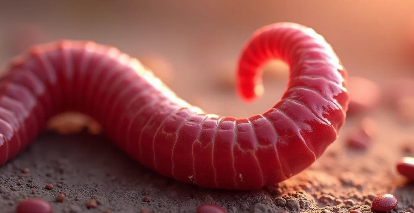
Ascaris lumbricoides, commonly known as the large roundworm, represents one of the most prevalent human parasitic infections globally. Adult worms can reach lengths of 15-35 centimetres and display a characteristic cylindrical shape with tapered ends. When expelled in stool, these worms may appear as elongated, root-like structures, particularly when multiple specimens become entangled or when partial segments are passed.
The distinctive morphology of Ascaris includes a smooth, cream-coloured cuticle that may appear branched when viewed superficially. Female worms are typically larger than males and possess a straight posterior end, while males exhibit a curved tail with copulatory spicules. The identification of these characteristics requires careful examination, as dead or partially digested worms may lose some defining features while retaining their basic root-like appearance.
Enterobius vermicularis Thread-Like appearance in faecal matter
Enterobius vermicularis, or pinworm, creates distinctly different presentations compared to larger roundworms. These small, white, thread-like parasites measure only 8-13 millimetres in length but often appear in clusters that can resemble fine root systems or fungal networks. The characteristic appearance results from multiple worms aggregating together, particularly around the perianal region where egg-laying occurs.
Pinworm identification relies heavily on their distinctive morphological features, including the pointed posterior end in females and the curved tail in males. When multiple specimens are present in stool samples, they may create complex branching patterns that closely resemble plant root systems. The thread-like consistency and white colouration distinguish pinworms from dietary fibres or other non-parasitic materials that might create similar appearances.
Trichuris trichiura Whip-Shaped structure recognition
Trichuris trichiura, known as whipworm, presents unique morphological characteristics that can contribute to root-like appearances in stool. The distinctive whip-shaped structure consists of a thin anterior end and a broader posterior portion, creating an overall appearance that may resemble branched plant material when multiple specimens are present or when viewed at certain angles.
Adult whipworms measure 30-50 millimetres in length and embed their thin anterior ends deep into the colonic mucosa. When expelled, particularly during treatment or natural elimination, these parasites may appear as complex, branching structures due to their unusual shape and tendency to become entangled with mucus and other intestinal contents. The recognition of whipworm infections requires understanding these distinctive morphological features and their potential to create misleading visual presentations.
Strongyloides stercoralis larval forms in fresh specimens
Strongyloides stercoralis presents particular diagnostic challenges due to its complex life cycle and the presence of larval forms in fresh faecal specimens. Unlike other parasitic worms that primarily appear as adults or eggs in stool, Strongyloides infections may produce motile larvae that create constantly changing, root-like patterns within fresh samples.
The larval forms of Strongyloides measure approximately 200-300 micrometres in length and display active motility when examined in fresh specimens. This movement can create the impression of branching, root-like structures as multiple larvae interact with surrounding material. The identification of Strongyloides requires prompt examination of fresh samples, as the characteristic larval motility diminishes rapidly after specimen collection.
Dietary fibre residue: cellulose and lignin fragment analysis
Plant-based dietary components represent the most common cause of root-like objects appearing in stool, with various fibrous materials creating convincing mimics of parasitic infections or other pathological conditions. The human digestive system cannot completely break down certain plant fibres, particularly cellulose and lignin structures, leading to the passage of recognisable plant fragments that maintain their original morphological characteristics.
Understanding the relationship between dietary intake and stool appearance requires knowledge of plant cell wall composition and digestive physiology. Cellulose, lignin, and other structural polysaccharides resist enzymatic breakdown in the human intestinal tract, allowing complex plant structures to pass through largely intact. These materials can aggregate with mucus and other intestinal contents to create elaborate, root-like formations that may cause significant concern.
Undigested vegetable matter from sweetcorn and bean consumption
Sweetcorn kernels and bean hulls represent classic examples of plant materials that create root-like appearances in stool. The fibrous outer layers of corn kernels, composed primarily of cellulose and lignin, resist complete digestion and may appear as yellow, branching structures when partially broken down. Similarly, bean hulls maintain their structural integrity through the digestive process, creating tough, root-like fragments.
The identification of vegetable-derived root-like objects relies on correlating recent dietary intake with stool appearances. Sweetcorn consumption typically results in recognisable yellow fragments within 24-48 hours of ingestion, while bean hulls may appear as brown or tan-coloured, fibrous material with characteristic branching patterns. These materials lack the uniform structure and biological characteristics of parasitic worms, helping distinguish dietary residues from infectious causes.
Fruit skin remnants from apple and grape ingestion
Fruit skins, particularly from apples, grapes, and stone fruits, contribute significantly to root-like appearances in stool due to their high concentrations of indigestible cellulose and waxy coatings. Apple peels contain complex networks of cellulose fibres reinforced with lignin, creating tough, branching structures that maintain their integrity throughout digestion.
Grape skins present unique challenges due to their combination of cellulose fibres and waxy cuticles that resist breakdown by digestive enzymes. When multiple grape skins aggregate in the intestinal tract, they can form complex, root-like masses that appear remarkably similar to parasitic infections. The identification of fruit-derived materials requires careful examination of colour, texture, and structural characteristics that distinguish plant matter from biological organisms.
Seed hull fragments from sesame and flaxseed sources
Small seeds, including sesame, flax, and poppy seeds, possess extremely durable hull structures designed to protect the internal embryo from environmental damage. These protective coatings, composed of multiple layers of cellulose and lignin, create some of the most persistent plant materials encountered in human digestion.
Flaxseed hulls demonstrate particular resistance to digestive breakdown due to their mucilaginous coating and tough cellulose structure. When consumed in quantity, these seeds may aggregate in the intestinal tract, forming complex, root-like masses that can cause significant concern. The identification of seed-derived materials relies on recognising their characteristic size, shape, and surface texture, which typically retain distinctive features even after partial digestion.
Nut shell particles from almond and walnut consumption
Tree nut shells represent some of the most durable plant materials encountered in human digestion, with almond and walnut shells creating particularly convincing root-like appearances when incompletely chewed or accidentally consumed. These materials consist of extremely dense lignin networks designed to protect developing nuts from environmental damage and predation.
Walnut shell fragments demonstrate remarkable durability, maintaining their characteristic brown colouration and fibrous texture throughout the digestive process. When multiple fragments aggregate with intestinal mucus, they can create elaborate branching patterns that closely resemble parasitic infections or other pathological conditions. The recognition of nut-derived materials requires understanding their distinctive hardness, colour patterns, and resistance to digestive breakdown.
Mucus cast formation: colonic and small intestinal origin
Intestinal mucus production serves essential protective and lubricating functions throughout the digestive tract, but certain pathological conditions can lead to excessive mucus formation that creates root-like appearances when expelled. Mucus casts form when excessive secretions aggregate with cellular debris, undigested food particles, and other intestinal contents to create complex, branching structures that may cause significant alarm.
The formation of mucus casts involves complex interactions between inflammatory processes, intestinal motility, and secretory function. Various disease states can trigger excessive mucus production, leading to the formation of elaborate casts that maintain branching patterns reflecting the internal architecture of the intestinal tract. Understanding these formations requires knowledge of both normal mucus physiology and the pathological processes that drive excessive secretion.
Mucus cast formation represents a significant diagnostic indicator of underlying intestinal inflammation or dysfunction, requiring careful evaluation to distinguish benign causes from serious pathological conditions.
Pseudomembranous colitis associated mucus strands
Pseudomembranous colitis, typically caused by Clostridioides difficile infection, produces characteristic mucus formations that can create convincing root-like appearances in stool. The inflammatory process triggers massive mucus secretion combined with epithelial cell shedding, creating complex pseudomembrane structures that may appear as branching, root-like objects when expelled.
The distinctive appearance of pseudomembranous colitis-related mucus casts includes yellow-green colouration, fibrin deposition, and cellular debris aggregation. These formations maintain structural integrity due to protein cross-linking and inflammatory mediator interactions, creating durable casts that closely resemble parasitic infections or plant materials. Recognition requires understanding the clinical context and associated symptoms of C. difficile infection.
Irritable bowel syndrome mucus production patterns
Irritable bowel syndrome (IBS) frequently involves altered mucus production patterns that can contribute to unusual stool appearances, including root-like formations. The condition affects intestinal motility and secretory function, leading to inconsistent mucus production that may aggregate into complex structures during certain symptom phases.
IBS-related mucus formations typically lack the inflammatory characteristics seen in infectious or inflammatory conditions but may still create convincing root-like appearances due to altered intestinal transit times and secretory patterns. The identification of IBS-related mucus requires understanding the chronic, relapsing nature of the condition and its relationship to stress, dietary factors, and hormonal influences.
Inflammatory bowel disease mucosal shedding
Inflammatory bowel disease, including Crohn’s disease and ulcerative colitis, produces significant mucosal inflammation and shedding that can create elaborate root-like formations in stool. The chronic inflammatory process leads to excessive mucus production combined with epithelial cell loss, creating complex aggregations that may appear as branching, plant-like structures.
The mucus casts associated with inflammatory bowel disease often contain blood, inflammatory cells, and necrotic tissue, creating distinctive colouration and texture patterns. These formations may demonstrate remarkable structural complexity due to the chronic nature of the inflammatory process and the extensive mucosal damage characteristic of these conditions. Recognition requires understanding the broader clinical presentation and associated symptoms of inflammatory bowel disease.
Foreign body ingestion: Non-Digestible material identification
Accidental or intentional ingestion of non-food materials can create surprising root-like appearances in stool, particularly when fibrous or branching materials pass through the digestive tract intact. Common sources include plant materials from outdoor activities, textile fibres from clothing or bedding, and various synthetic materials encountered in daily life.
Paper products, including tissue paper and cardboard fragments, can create convincing root-like appearances when partially broken down by digestive processes. The cellulose fibres in these materials maintain structural integrity while becoming softened and aggregated with intestinal contents. Hair and textile fibres may also contribute to unusual stool appearances, particularly when consumed in quantity or when digestive transit times are prolonged.
Foreign body identification requires careful consideration of recent activities, environmental exposures, and the possibility of pica or other behavioural factors that might lead to non-food ingestion.
Pharmaceutical excipient remnants: Modified-Release tablet matrices
Modern pharmaceutical formulations often employ complex excipient systems designed to control drug release and protect active ingredients during gastrointestinal transit. These excipients, particularly in extended-release and enteric-coated formulations, may create unusual appearances in stool that can resemble root-like structures when expelled intact or partially dissolved.
Hydroxypropyl methylcellulose (HPMC) matrices, commonly used in sustained-release tablets, can maintain structural integrity throughout much of the digestive process. When these materials aggregate with intestinal contents, they may create gel-like, branching formations that closely resemble biological materials. The identification of pharmaceutical-derived materials requires correlating medication use with stool appearance timing and understanding the specific excipient systems employed in individual formulations.
Pharmaceutical excipients designed for controlled drug release may create surprisingly durable structures that maintain recognisable forms throughout gastrointestinal transit, potentially causing diagnostic confusion.
Diagnostic laboratory testing: microscopic examination and parasitological methods
Accurate identification of root-like objects in stool requires systematic laboratory examination employing multiple diagnostic techniques. Fresh specimen collection and prompt analysis provide optimal conditions for identifying motile parasites, while preserved samples allow for detailed morphological examination and special staining procedures.
Direct microscopic examination remains the cornerstone of parasitological diagnosis, utilising both wet mount preparations and permanent stained slides to identify characteristic morphological features. Concentration techniques, including formalin-ethyl acetate sedimentation and zinc sulphate flotation, enhance parasite detection sensitivity by removing debris and concentrating organisms. Modern molecular diagnostic methods, including PCR-based assays, provide additional confirmation for suspected parasitic infections while offering improved sensitivity and specificity compared to traditional microscopy.
The interpretation of laboratory results requires understanding the limitations of different diagnostic techniques and the importance of clinical correlation. False-positive results may occur when plant materials or other debris mimic parasitic structures, while false-negative results can result from intermittent parasite shedding or inappropriate specimen collection and handling. Experienced laboratory personnel play crucial roles in distinguishing genuine parasitic infections from benign dietary or pharmaceutical residues that create similar appearances.