The anatomy beneath the right rib cage houses several vital organs that perform essential functions for human survival. The liver dominates this region, occupying the largest portion of the right upper quadrant alongside the gallbladder, right kidney, and portions of the ascending colon. Understanding the precise anatomical arrangement of these structures proves crucial for healthcare professionals diagnosing abdominal pain and for individuals seeking to comprehend their body’s complex organisation. The intricate positioning of these organs creates a sophisticated system where each structure works in harmony with neighbouring tissues to maintain optimal physiological function.
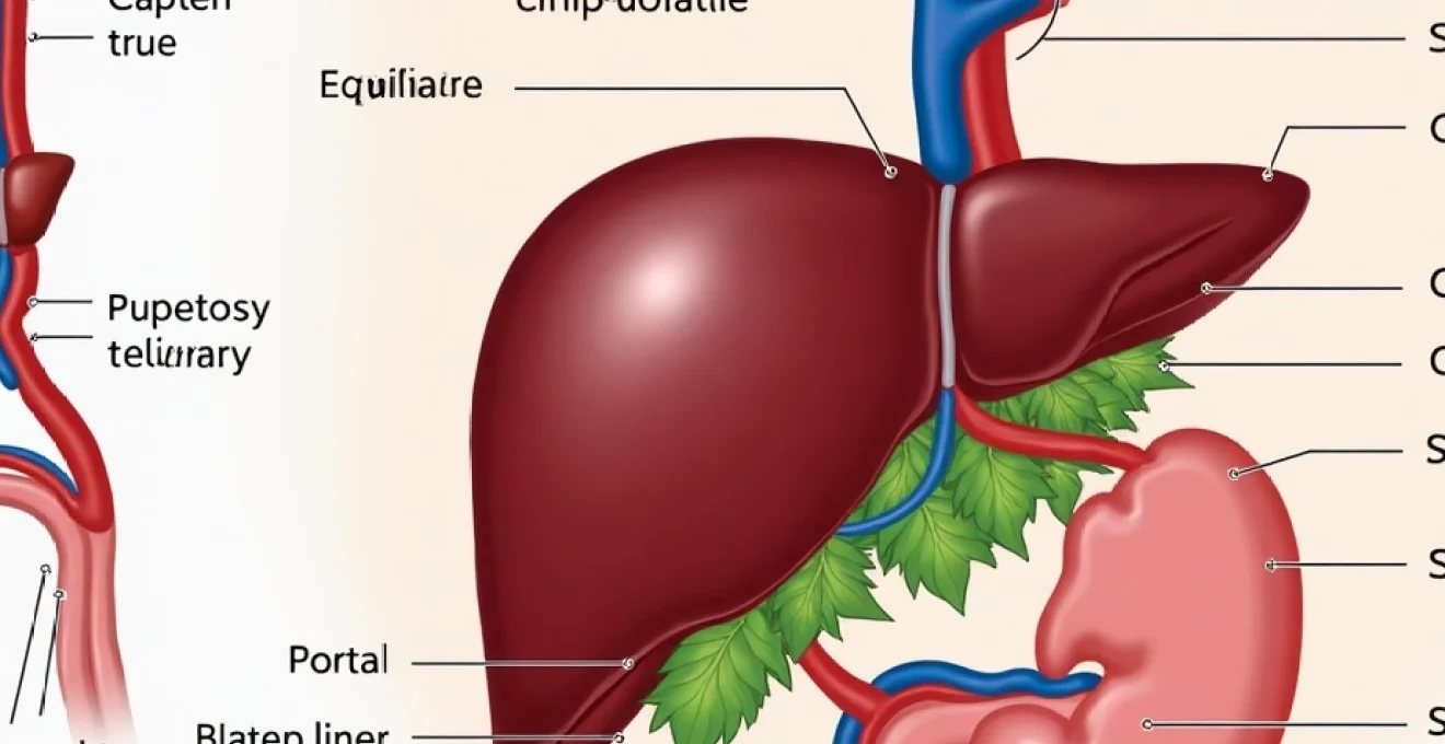
Anatomical structure of the liver in the right hypochondriac region
The liver represents the largest internal organ within the human body, weighing approximately 1.5 kilograms in healthy adults. This remarkable structure extends across both the right and left upper quadrants, though its dominant mass resides beneath the right costal margin. The liver’s strategic positioning allows it to perform over 500 distinct metabolic functions whilst maintaining direct anatomical relationships with surrounding viscera.
Hepatic lobar architecture and couinaud segmental classification
The liver’s complex architecture divides into two primary lobes separated by the falciform ligament. The right lobe significantly exceeds the left lobe in size, comprising approximately 60% of the organ’s total mass. Modern hepatic surgery relies upon the Couinaud classification system, which delineates the liver into eight functional segments based on portal vein distribution patterns. This segmental approach enables precise surgical planning and allows surgeons to perform targeted resections whilst preserving maximum hepatic function.
Each hepatic segment receives independent blood supply from portal vein branches and hepatic artery divisions. The anatomical significance of this arrangement becomes apparent during pathological processes, where disease may affect specific segments whilst sparing adjacent areas. Segmental anatomy proves particularly relevant when evaluating hepatocellular carcinoma distribution or planning living donor liver transplantation procedures.
Portal triad components and glisson’s capsule anatomy
The portal triad represents a fundamental structural unit comprising the hepatic artery, portal vein, and bile duct enclosed within Glisson’s capsule. This fibrous sheath extends throughout the hepatic parenchyma, creating a supportive framework for vascular and biliary structures. The portal vein contributes approximately 75% of the liver’s blood supply, whilst the hepatic artery provides the remaining 25% under normal physiological conditions.
Glisson’s capsule demonstrates remarkable tensile strength, protecting delicate hepatic structures from external trauma. During hepatic inflammation or congestion, capsular distension creates the characteristic right upper quadrant discomfort experienced by patients with acute hepatitis or congestive heart failure. The capsule’s innervation by sympathetic fibres explains the referred pain patterns commonly observed in hepatic pathology.
Hepatocyte microstructure and sinusoidal blood flow patterns
Hepatocytes arrange themselves in organised plates radiating from central veins toward portal triads, creating the classic hepatic lobule architecture. These specialised cells demonstrate remarkable regenerative capacity, enabling the liver to restore functional mass following injury or surgical resection. Hepatic sinusoids facilitate efficient blood-hepatocyte contact, maximising metabolic exchange and detoxification processes.
Kupffer cells line the sinusoidal spaces, serving as the liver’s resident macrophage population. These immune cells play crucial roles in bacterial clearance and inflammatory responses. The sinusoidal endothelium maintains unique fenestrations that allow free passage of plasma proteins whilst restricting cellular elements, creating an optimal environment for hepatic metabolic functions.
Falciform ligament and coronary ligament attachments
The falciform ligament serves as the primary anterior hepatic attachment, extending from the diaphragm to the umbilicus whilst dividing the liver into anatomical right and left lobes. This peritoneal fold contains the ligamentum teres, a remnant of the fetal umbilical vein that occasionally remains patent in certain pathological conditions. The coronary ligaments provide posterior hepatic fixation, creating bare areas where the liver directly contacts the diaphragmatic surface.
These ligamentous attachments prove clinically significant during hepatic mobilisation procedures and laparoscopic surgery. Understanding their anatomy prevents inadvertent injury to surrounding structures and enables safe hepatic manipulation. The triangular ligaments represent lateral extensions of the coronary ligaments, providing additional stabilisation for the hepatic margins.
Gallbladder positioning and biliary tree configuration
The gallbladder occupies a strategic position within the hepatocystic fossa, nestled beneath the liver’s quadrate lobe. This pear-shaped organ measures approximately 7-10 centimetres in length and maintains capacity for 30-50 millilitres of concentrated bile. The gallbladder’s anatomical relationships prove crucial for understanding biliary pathophysiology and surgical approaches to cholecystic disease.
Fundus, body, and neck anatomical landmarks
The gallbladder’s fundus projects beyond the hepatic margin, creating a palpable structure during physical examination when pathologically distended. The body represents the gallbladder’s largest portion, whilst the neck tapers toward the cystic duct junction. Anatomical variations in gallbladder morphology occur frequently, including septations, duplications, and aberrant positioning that may complicate surgical procedures.
The gallbladder wall comprises four distinct layers: mucosa, muscularis, perimuscular connective tissue, and serosa. The mucosal surface demonstrates characteristic rugal folds that flatten during distension. Rokitansky-Aschoff sinuses represent mucosal invaginations that may become inflamed during chronic cholecystitis, creating diagnostic challenges on imaging studies.
Cystic artery variations and calot’s triangle boundaries
Calot’s triangle defines the critical anatomical space bounded by the cystic artery, common hepatic duct, and cystic duct. This region contains the cystic artery in approximately 95% of individuals, though significant anatomical variations exist. The cystic artery typically arises from the right hepatic artery, though aberrant origins from the left hepatic artery, gastroduodenal artery, or superior mesenteric artery occur in 15-20% of cases.
Understanding cystic arterial anatomy proves essential for safe cholecystectomy procedures. Accessory cystic arteries may supply the gallbladder neck or hepatocystic triangle, requiring careful identification during surgical dissection. The hepatocystic artery occasionally provides additional blood supply to the gallbladder fundus, particularly in elderly patients with atherosclerotic disease.
Common bile duct pathway through ampulla of vater
The common bile duct traverses a complex anatomical pathway from the hepatic confluence to the duodenal ampulla. This structure measures 6-8 centimetres in length and demonstrates variable diameter depending on age and pathological conditions. The duct’s intrapancreatic segment proves particularly susceptible to compression from pancreatic pathology or periampullary tumours.
The sphincter of Oddi regulates bile flow into the duodenum through coordinated muscular contractions. This sophisticated mechanism prevents duodenal contents from refluxing into the biliary tree whilst allowing coordinated bile and pancreatic juice delivery during digestion. Sphincter dysfunction may produce recurrent biliary-type pain even following cholecystectomy.
Hartmann’s pouch and Rokitansky-Aschoff sinus formation
Hartmann’s pouch represents a common anatomical variant where the gallbladder neck demonstrates outpouching adjacent to the cystic duct junction. This configuration predisposes to stone impaction and may complicate endoscopic interventions. The pouch’s relationship to surrounding structures requires careful consideration during laparoscopic cholecystectomy to prevent ductal injury.
Rokitansky-Aschoff sinuses develop through chronic inflammatory processes that create mucosal herniation through the gallbladder wall. These pseudodiverticular formations may harbour bacteria and contribute to recurrent cholecystitis episodes. Advanced imaging techniques increasingly identify these structures, influencing surgical planning and patient counselling regarding long-term outcomes.
Right kidney and adrenal gland retroperitoneal location
The right kidney occupies the retroperitoneal space posterior to the ascending colon and hepatic flexure. This bean-shaped organ measures approximately 10-12 centimetres in length and maintains close anatomical relationships with the liver, duodenum, and right colic vessels. The kidney’s position beneath the right costal margin explains why renal pathology frequently presents with flank pain radiating to the subcostal region.
The right kidney typically sits slightly lower than its left counterpart due to hepatic displacement. This anatomical difference proves clinically significant when interpreting imaging studies or performing percutaneous interventions. The renal fascia creates distinct compartments that influence the spread of pathological processes and guide surgical approaches to retroperitoneal structures.
The right adrenal gland caps the superior renal pole, demonstrating a triangular configuration that differs from the left adrenal’s crescentic shape. This endocrine organ produces essential hormones including cortisol, aldosterone, and catecholamines. Adrenal pathology may present with right upper quadrant discomfort due to capsular distension or adjacent organ compression. The adrenal’s rich vascular supply includes contributions from the superior, middle, and inferior adrenal arteries, creating potential haemorrhagic complications during surgical manipulation.
Hepatic flexure of the colon and ascending bowel segment
The hepatic flexure represents the junction between the ascending and transverse colon, positioned directly beneath the liver’s inferior surface. This anatomical landmark proves crucial for colonoscopic navigation and surgical planning during colectomy procedures. The flexure’s acute angulation may trap gas bubbles, creating the characteristic hepatic flexure syndrome with associated right upper quadrant discomfort.
The ascending colon traverses the right paracolic gutter from the caecum to the hepatic flexure, maintaining retroperitoneal positioning throughout its course. This segment demonstrates variable mobility depending on developmental variations in peritoneal attachments. Some individuals exhibit a mobile ascending colon with a mesentery, whilst others demonstrate fixed retroperitoneal positioning that influences surgical approaches and complication rates.
Vascular supply to the ascending colon derives primarily from the ileocolic and right colic arteries, branches of the superior mesenteric arterial system. Understanding this vascular anatomy proves essential for maintaining adequate perfusion during surgical resection. The marginal artery of Drummond provides collateral circulation that may prevent ischaemic complications when primary vessels are compromised. Venous drainage follows arterial patterns, ultimately reaching the portal circulation through the superior mesenteric vein.
Pathological conditions affecting right subcostal organs
Numerous pathological processes may affect organs beneath the right rib cage, creating diagnostic challenges for healthcare providers. The overlapping innervation patterns and shared anatomical relationships between these structures often produce similar clinical presentations despite diverse underlying aetiologies. Understanding the characteristic features of common right upper quadrant pathologies enables accurate diagnosis and appropriate therapeutic interventions.
Hepatomegaly secondary to cirrhosis and fatty liver disease
Hepatomegaly represents a common physical finding in patients with chronic liver disease, creating palpable enlargement below the right costal margin. Cirrhosis initially produces hepatomegaly through inflammatory processes and regenerative nodule formation, though end-stage disease may result in hepatic atrophy. Non-alcoholic fatty liver disease (NAFLD) has emerged as the leading cause of hepatomegaly in developed nations, affecting up to 25% of the adult population.
The clinical presentation of hepatomegaly depends upon the underlying aetiology and disease progression. Patients may experience right upper quadrant fullness , early satiety, or referred shoulder pain due to diaphragmatic irritation. Physical examination reveals a palpable liver edge extending below the costal margin, though percussion techniques provide more reliable assessment of hepatic size and consistency.
Imaging studies demonstrate characteristic patterns associated with different hepatic pathologies. Ultrasound reveals increased echogenicity in fatty infiltration, whilst cirrhosis produces a nodular contour with surface irregularity. Advanced fibrosis assessment utilises elastography techniques that measure hepatic stiffness, providing non-invasive alternatives to liver biopsy for staging chronic liver disease.
Acute cholecystitis and cholelithiasis clinical presentations
Acute cholecystitis typically presents with sudden-onset right upper quadrant pain that may radiate to the epigastrium or right shoulder blade. The pathophysiology involves cystic duct obstruction leading to gallbladder distension, inflammation, and potential secondary bacterial infection. Murphy’s sign represents a classic physical finding where inspiration during right subcostal palpation produces pain and respiratory arrest.
Cholelithiasis affects approximately 10-15% of adults in Western populations, with prevalence increasing significantly with age. The majority of gallstones remain asymptomatic throughout an individual’s lifetime, though complications may develop when stones obstruct the cystic or common bile ducts. Biliary colic produces characteristic episodic pain that builds to a crescendo over 15-30 minutes before gradually subsiding.
Gallstone complications include acute cholecystitis, choledocholithiasis, cholangitis, and gallstone pancreatitis, each requiring specific therapeutic approaches and timeline considerations.
The differential diagnosis of right upper quadrant pain extends beyond simple cholelithiasis to include acalculous cholecystitis, gallbladder polyps, and cholangiocarcinoma. Advanced age, diabetes, and immunocompromised states predispose to acalculous cholecystitis, which demonstrates higher morbidity and mortality rates compared to calculous disease. Prompt recognition and appropriate intervention prove crucial for optimal patient outcomes.
Right-sided nephrolithiasis and hydronephrosis complications
Renal calculi affecting the right kidney commonly present with severe flank pain that may radiate to the groin or lower abdomen. The pain typically demonstrates sudden onset and may be accompanied by nausea, vomiting, and haematuria. Stone composition varies geographically and demographically, with calcium oxalate representing the most common variety in developed nations.
Hydronephrosis develops when urinary obstruction produces renal pelvicalyceal dilatation and potential parenchymal damage. Acute obstruction creates severe pain and requires urgent intervention to preserve renal function. Chronic hydronephrosis may remain asymptomatic until significant functional impairment occurs, emphasising the importance of regular monitoring in high-risk populations.
Imaging plays a crucial role in nephrolithiasis diagnosis and management planning. Non-contrast computed tomography demonstrates superior sensitivity for stone detection compared to traditional intravenous pyelography. Ultrasound provides valuable information regarding hydronephrosis severity and may identify radiolucent stones missed by plain radiography. Ureteral stenting or percutaneous nephrostomy may be required for acute obstructive complications whilst definitive stone treatment is planned.
Diagnostic imaging techniques for right upper quadrant assessment
Modern imaging technologies provide sophisticated tools for evaluating right upper quadrant pathology with exceptional accuracy and detail. The selection of appropriate imaging modalities depends upon clinical presentation, suspected pathology, and patient-specific factors including renal function and contrast allergies. Multi-modal imaging approaches often prove necessary for comprehensive assessment of complex abdominal conditions.
Ultrasound murphy’s sign and gallbladder wall thickening
Ultrasonographic Murphy’s sign represents a highly specific finding in acute cholecystitis, demonstrating superior diagnostic accuracy compared to clinical examination alone. This technique involves applying transducer pressure directly over the sonographically identified gallbladder whilst observing patient response. Positive findings include focal tenderness and involuntary respiratory arrest during inspiration, correlating strongly with cystic duct obstruction.
Gallbladder wall thickening exceeding 3 millimetres suggests acute inflammatory processes, though this finding lacks specificity for cholecystitis. Alternative causes include hepatitis, congestive heart failure, hypoproteinaemia, and chronic kidney disease. The combination of wall thickening, pericholecystic fluid, and positive sonographic Murphy’s sign provides diagnostic accuracy exceeding 95
% for acute cholecystitis diagnosis. Additional ultrasonographic features include gallbladder distension, echogenic bile, and the presence of gallstones with posterior acoustic shadowing.
Real-time ultrasound assessment enables dynamic evaluation of gallbladder contractility and bile duct dilatation. The hepatocystic triangle requires careful examination to identify inflammatory changes or anatomical variants that may complicate surgical intervention. Doppler ultrasound techniques demonstrate increased vascularity within thickened gallbladder walls, supporting the diagnosis of acute inflammatory processes.
CT hepatobiliary phase enhancement patterns
Multiphasic computed tomography provides comprehensive evaluation of hepatobiliary pathology through carefully timed contrast administration. The hepatobiliary phase, obtained 15-20 minutes following contrast injection, demonstrates optimal parenchymal enhancement patterns that reveal structural abnormalities and functional impairments. Hepatocyte-specific contrast agents such as gadoxetic acid enable simultaneous assessment of liver function and biliary excretion.
Normal hepatic enhancement demonstrates homogeneous attenuation throughout all hepatic segments, with portal and hepatic veins clearly delineated. Pathological conditions produce characteristic enhancement patterns: hypervascular lesions enhance brightly during arterial phase imaging, whilst hypovascular lesions remain relatively hypodense. Delayed phase imaging proves particularly valuable for characterising haemangiomas and identifying small hepatocellular carcinomas.
Biliary tree assessment utilises thin-section reconstructions that demonstrate both intra- and extrahepatic ductal anatomy. Choledocholithiasis appears as filling defects within contrast-enhanced bile ducts, whilst strictures produce focal narrowing with upstream dilatation. The combination of parenchymal and ductal imaging enables comprehensive evaluation of complex hepatobiliary pathology requiring multidisciplinary management approaches.
MRCP visualisation of biliary tree obstruction
Magnetic resonance cholangiopancreatography (MRCP) represents the gold standard non-invasive technique for biliary tree assessment. This technology utilises heavily T2-weighted sequences that highlight fluid-filled structures whilst suppressing background tissues. The resulting images provide exquisite detail of biliary anatomy without requiring contrast administration or invasive procedures.
MRCP demonstrates superior sensitivity for detecting choledocholithiasis compared to ultrasound or computed tomography. Small stones measuring 2-3 millimetres appear as signal voids within high-intensity bile, enabling detection of clinically significant obstruction before symptoms develop. Biliary strictures produce characteristic focal narrowing with associated upstream dilatation, facilitating differentiation between benign and malignant aetiologies.
Three-dimensional MRCP reconstructions provide comprehensive visualisation of complex biliary anatomy and variant configurations. These techniques prove particularly valuable for preoperative planning in patients requiring hepatobiliary surgery or endoscopic intervention. The combination of MRCP with conventional MRI sequences enables simultaneous assessment of biliary pathology and surrounding soft tissue abnormalities.
Advanced MRCP techniques including secretin stimulation and pharmacological enhancement provide functional information regarding sphincter of Oddi function and pancreatic duct compliance.
HIDA scan functional gallbladder assessment
Hepatobiliary iminodiacetic acid (HIDA) scintigraphy provides unique functional assessment of biliary excretion and gallbladder contractility. This nuclear medicine technique utilises technetium-99m labelled compounds that undergo hepatic uptake and biliary excretion, enabling real-time visualisation of bile flow dynamics. The study proves particularly valuable when anatomical imaging techniques demonstrate equivocal findings.
Normal HIDA scans demonstrate prompt hepatic uptake within 5-10 minutes, followed by biliary excretion and gallbladder filling within 30-60 minutes. Acute cholecystitis produces characteristic non-visualisation of the gallbladder despite normal hepatic uptake and bile duct filling. Chronic cholecystitis may demonstrate delayed gallbladder filling or impaired contractility following cholecystokinin stimulation.
Quantitative gallbladder ejection fraction assessment utilises standardised cholecystokinin administration to evaluate contractile function. Normal ejection fractions exceed 35-40%, whilst values below this threshold suggest gallbladder dyskinesia or chronic inflammatory changes. Functional biliary disorders may produce typical biliary pain despite normal anatomical imaging, making HIDA scanning essential for comprehensive evaluation of suspected gallbladder pathology.
The integration of functional and anatomical imaging techniques enables precise characterisation of right upper quadrant pathology. Understanding the capabilities and limitations of each modality ensures appropriate test selection and interpretation, ultimately facilitating optimal patient care and therapeutic outcomes. Advanced imaging technologies continue evolving, providing increasingly sophisticated tools for evaluating the complex anatomical relationships beneath the right rib cage.