Experiencing a bulky sensation with your new dental bridge can be both uncomfortable and concerning. This common post-treatment issue affects many patients, often causing difficulty with speech, chewing, and overall oral comfort. While some initial adjustment period is normal after bridge placement, persistent bulkiness may indicate underlying problems that require professional attention. Understanding the causes, assessment methods, and available solutions can help you address this issue effectively and restore your oral comfort.
The sensation of bulkiness in dental bridges typically stems from various anatomical and technical factors. Modern dentistry has developed sophisticated approaches to diagnose and treat these concerns, ensuring patients achieve optimal comfort and functionality. Whether the issue requires minor adjustments or more comprehensive intervention depends on the specific underlying causes and the severity of the problem.
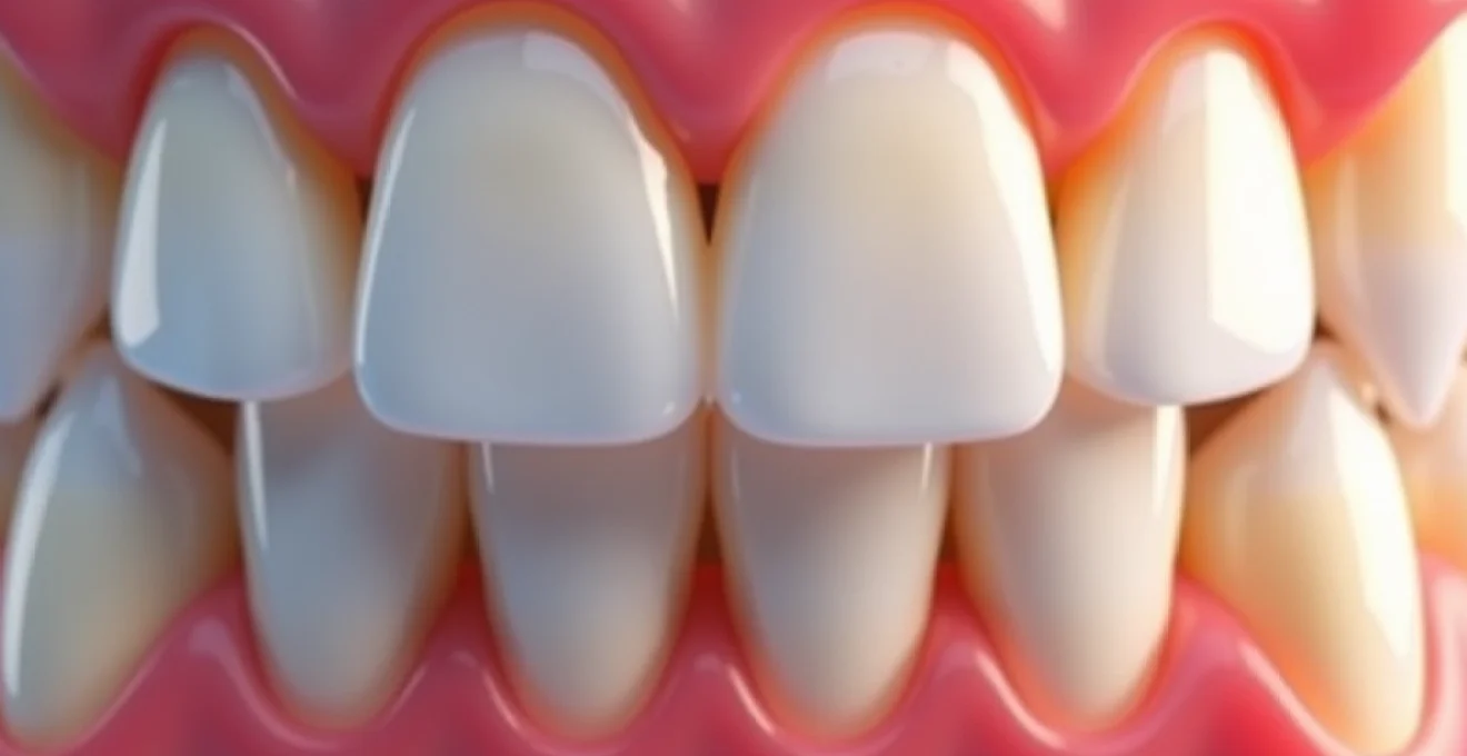
Understanding bulky dental bridge sensations and anatomical factors
The perception of bulkiness in dental bridges often relates to complex anatomical changes that occur following tooth loss and subsequent restoration. Your mouth undergoes significant adaptations when natural teeth are replaced with prosthetic alternatives, and these changes can create unfamiliar sensations that may persist beyond the typical adjustment period.
Occlusal vertical dimension changes after bridge cementation
Bridge placement frequently alters the occlusal vertical dimension , which refers to the space between your upper and lower teeth when your jaw is in a resting position. When this dimension increases even slightly, you may experience a feeling of thickness or bulkiness in your bite. This change occurs because the bridge’s height may differ from your natural teeth, requiring your jaw muscles to adapt to a new resting position. The adjustment process can take several weeks, during which time the bulky sensation may gradually diminish as your neuromuscular system adapts to the new dimensions.
Gingival contour alterations around pontic design
The artificial tooth portion of your bridge, known as the pontic, must be designed to maintain proper gum health while providing adequate function. However, gingival contour alterations around the pontic can create sensations of bulkiness, particularly if the restoration doesn’t follow the natural tissue architecture. The pontic’s emergence profile and contact with the underlying gum tissue play crucial roles in both comfort and cleanliness. When these elements are not optimally designed, patients often report feeling as though food traps more easily or that the bridge feels unnaturally thick in certain areas.
Tongue space reduction and lingual flange positioning
Your tongue requires adequate space to function properly during speech and swallowing. Bridge restorations that encroach upon this tongue space can create persistent feelings of bulkiness, particularly on the lingual (tongue-side) surfaces. The positioning of the lingual flange—the part of the bridge that faces your tongue—significantly impacts your comfort level. Even minor discrepancies in this area can result in chronic irritation and the sensation that something foreign occupies your mouth. Proper lingual contours should closely mimic the natural tooth anatomy to maintain comfortable tongue function.
Interdental papilla displacement effects
The small triangular pieces of gum tissue between your teeth, called interdental papillae, can become displaced or compressed during bridge placement. This displacement often results in altered sensations around the restoration, contributing to the overall feeling of bulkiness. When papillae don’t properly fill the spaces around your bridge, you may notice changes in how food feels during chewing or experience increased awareness of the restoration’s presence. These effects can be particularly pronounced in the anterior region where aesthetic and functional demands are highest.
Clinical assessment techniques for bridge bulkiness evaluation
Professional evaluation of bridge bulkiness requires systematic assessment using various clinical techniques. Modern dental practices employ multiple diagnostic approaches to identify specific areas of concern and develop targeted treatment plans. Understanding these assessment methods can help you communicate more effectively with your dental team about your specific symptoms and concerns.
Articulating paper analysis for high contact points
Articulating paper analysis represents one of the most fundamental diagnostic tools for evaluating bridge contact issues. This technique involves placing thin coloured paper between your teeth while you bite down, revealing areas of excessive contact that may contribute to bulkiness sensations. High contact points often create feelings of thickness or interference during normal jaw function. Your dentist will systematically evaluate these contact patterns in various jaw positions, including centric occlusion, lateral movements, and protrusive positions. The resulting marks on the paper provide a visual map of pressure distribution, allowing for precise identification of areas requiring adjustment.
Periodontal probing depth measurements around abutments
Periodontal health assessment around bridge abutments provides crucial information about tissue adaptation and potential sources of discomfort. Increased probing depths may indicate areas where the bridge margins are creating tissue irritation, contributing to bulkiness sensations. Your dental professional will carefully measure pocket depths around each abutment tooth, comparing these measurements to baseline values established before treatment. Significant increases in probing depths often correlate with overcontoured bridge margins that compress gum tissue and create uncomfortable sensations.
Radiographic assessment using bitewing and periapical films
Radiographic evaluation provides essential information about the bridge’s relationship to surrounding structures that cannot be assessed clinically. Bitewing radiographs reveal the crown-to-root ratio of abutment teeth and can identify areas where bridge contours may be contributing to food retention or tissue irritation. Periapical films offer detailed views of the entire root structure and surrounding bone, helping identify any underlying pathology that might influence your perception of bridge bulkiness. These images also allow assessment of margin adaptation and cement washout, both factors that can affect comfort and function.
Digital occlusal analysis with T-Scan technology
Advanced digital occlusal analysis systems like T-Scan technology provide precise, objective measurements of bite force distribution and timing. This sophisticated approach goes beyond traditional articulating paper analysis by measuring both the force and temporal aspects of occlusal contact. The system generates detailed maps showing exactly where and when your teeth contact during closing, revealing subtle timing discrepancies that may contribute to bulkiness sensations. This technology is particularly valuable for complex cases where traditional assessment methods may not fully capture the nature of the occlusal problem.
Professional adjustment procedures for oversized bridge restorations
When clinical assessment confirms that your bridge requires modification, several professional adjustment procedures can address bulkiness effectively. These techniques require specialized training and equipment to ensure precise modifications without compromising the restoration’s integrity or longevity. Understanding these procedures can help you make informed decisions about your treatment options.
Selective occlusal grinding with diamond burs
Selective occlusal adjustment using diamond burs represents the most common approach to reducing bridge bulkiness. This precise technique involves carefully removing small amounts of material from specific contact points identified during clinical assessment. Your dentist will use various diamond bur sizes and shapes to refine the occlusal anatomy while maintaining proper contact relationships with adjacent and opposing teeth. The procedure requires careful attention to cusp-fossa relationships and guidance patterns to ensure optimal function after adjustment. Multiple appointments may be necessary to achieve the ideal balance between comfort and functionality.
Pontic recontouring using coarse and fine polishing discs
Pontic recontouring addresses bulkiness in the artificial tooth portion of your bridge, particularly focusing on areas that contact the underlying gum tissue. This process typically begins with coarse polishing discs to remove larger amounts of material, followed by progressively finer discs to achieve smooth, biocompatible surfaces. The recontouring process must maintain adequate strength in the pontic while creating more natural contours that feel comfortable against your tongue and cheek tissues. Proper pontic design should allow for easy cleaning while minimizing food retention and tissue irritation.
Lingual surface reduction techniques
Lingual surface reduction specifically addresses tongue space concerns by carefully removing material from the inner aspects of your bridge. This delicate procedure requires precise understanding of tongue function patterns and speech requirements. Your dentist will systematically reduce bulky areas while maintaining structural integrity and ensuring adequate material thickness for long-term durability. The process often involves multiple small adjustments followed by careful polishing to achieve smooth, comfortable surfaces that allow normal tongue movement during speech and swallowing.
Embrasure space enhancement methods
Enhancing embrasure spaces—the triangular spaces between teeth—can significantly improve the comfort and cleanliness of your bridge. These spaces must be properly shaped to allow adequate access for cleaning while providing natural-feeling contours. Enhancement techniques involve carefully opening and reshaping these areas using specialized instruments and rotary tools. The goal is to create embrasure forms that closely match natural tooth anatomy while facilitating effective plaque removal and reducing food impaction that can contribute to bulkiness sensations.
Surface refinishing with astropol and enhance systems
After any adjustment procedure, surface refinishing becomes critical for both comfort and longevity. Advanced polishing systems like Astropol and Enhance provide systematic approaches to achieving optimal surface smoothness and lustre. These multi-step systems progress from coarser to finer abrasives, ultimately creating surfaces that resist plaque accumulation and feel comfortable against oral tissues. Proper surface finishing also helps prevent bacterial colonization and staining, contributing to both the health and aesthetic appearance of your restored bridge.
Immediate relief strategies for bridge discomfort management
While professional adjustment remains the definitive solution for bulky bridge sensations, several immediate relief strategies can help manage discomfort during the interim period. These approaches focus on reducing inflammation, managing sensitivity, and optimizing your oral environment to minimize irritation until professional treatment can be completed.
Soft tissue management becomes particularly important when dealing with bridge-related bulkiness. Gentle massage of the gum tissues around your bridge can help improve circulation and reduce localized swelling that may contribute to discomfort sensations. Using your clean fingertip, apply light circular pressure to the gum areas adjacent to the bridge for several minutes, twice daily. This simple technique can help tissues adapt more quickly to the new restoration while reducing inflammatory responses that may amplify bulkiness perceptions.
Dietary modifications play a crucial role in immediate comfort management. Focus on softer foods that require minimal chewing force and avoid particularly chewy or sticky items that may increase your awareness of the bridge’s presence. Temperature considerations also matter—extremely hot or cold foods may heighten sensitivity and make bulkiness sensations more pronounced. Room temperature or lukewarm foods often provide the most comfortable eating experience during the adjustment period.
Oral hygiene adaptations can significantly impact your comfort level while managing bridge bulkiness. Traditional flossing may feel more challenging with a bulky restoration, so consider using floss threaders or water flossers to maintain cleanliness without creating additional pressure on sensitive tissues. Gentle brushing with an extra-soft toothbrush can help maintain oral health without aggravating already sensitive areas around your bridge.
Professional intervention remains essential for addressing persistent bridge bulkiness, but immediate relief strategies can provide temporary comfort and facilitate better tissue adaptation during the adjustment period.
Long-term solutions and prosthetic replacement considerations
When adjustment procedures fail to adequately address bridge bulkiness, or when the restoration exhibits fundamental design flaws, replacement may represent the most effective long-term solution. Modern prosthetic replacement approaches offer significant advantages over traditional methods, incorporating advanced materials and digital design techniques that can dramatically improve both comfort and function.
Contemporary bridge replacement often involves digital impression techniques that capture more precise anatomical details than traditional impression materials. These digital workflows allow for more accurate recreation of your natural tooth contours and better integration with existing oral structures. Computer-aided design and manufacturing (CAD/CAM) technologies enable precise control over bridge dimensions and contours, significantly reducing the likelihood of bulkiness issues in the replacement restoration.
Material selection plays a crucial role in long-term comfort and function. Advanced ceramic materials offer superior biocompatibility and can be fabricated with thinner cross-sections while maintaining adequate strength. This characteristic allows for more conservative tooth preparation and less bulky final restorations. Zirconia-based materials, in particular, provide excellent strength-to-thickness ratios, enabling the creation of bridges that feel more natural while providing excellent longevity.
The decision between repair and replacement requires careful consideration of multiple factors, including the age of the existing restoration, the extent of required modifications, and your individual anatomy. Extensive adjustments may compromise the structural integrity of older bridges, making replacement a more predictable option. Additionally, bridges showing signs of wear, discoloration, or marginal breakdown may benefit from complete replacement rather than continued repair attempts.
Long-term success with bridge restorations depends heavily on proper initial design and fabrication, emphasizing the importance of working with experienced dental professionals who understand the complexities of prosthetic comfort and function.
Prevention protocols for future bridge fabrication accuracy
Preventing bulkiness issues in future bridge work requires comprehensive planning and meticulous attention to detail throughout the treatment process. Understanding these prevention protocols can help you advocate for optimal treatment approaches and collaborate more effectively with your dental team to achieve superior results.
Pre-treatment planning represents the foundation of successful bridge therapy. Comprehensive examination including detailed periodontal assessment, occlusal analysis, and radiographic evaluation helps identify potential challenges before treatment begins. Diagnostic wax-ups and provisional restorations provide valuable opportunities to test proposed dimensions and contours before final fabrication, allowing modifications that prevent bulkiness issues in the definitive restoration.
Communication protocols between you and your dental team significantly impact treatment outcomes. Detailed discussions about your expectations, concerns, and any history of sensitivity to previous restorations help guide treatment planning decisions. Regular feedback during provisional restoration phases allows for refinements that can be incorporated into the final bridge design, reducing the likelihood of post-cementation adjustment requirements.
Laboratory collaboration plays an essential role in achieving optimal bridge contours and dimensions. Advanced dental laboratories employ skilled technicians who understand the subtle anatomical variations that influence patient comfort. Providing detailed clinical information, including photographs and precise shade matching, helps laboratory technicians create restorations that integrate seamlessly with your existing oral structures.
Quality control measures throughout the fabrication process help ensure dimensional accuracy and proper fit. This includes careful evaluation of laboratory work before cementation, try-in procedures that allow final adjustments, and systematic verification of occlusal relationships. Modern digital verification techniques can identify potential problems before final cementation, allowing corrections that prevent post-treatment bulkiness issues.