Blood collection procedures, whilst generally safe and routine, can occasionally trigger unexpected dermatological reactions that range from mild irritation to severe allergic responses. These post-venipuncture skin manifestations affect approximately 2-5% of patients undergoing phlebotomy, with reactions varying significantly in severity, onset time, and clinical presentation. Understanding the underlying mechanisms behind these cutaneous responses is crucial for healthcare professionals and patients alike, as proper identification can prevent complications and guide appropriate treatment strategies.
The development of rashes following blood draws represents a complex interplay of immunological, mechanical, and chemical factors that can manifest immediately or develop over several days. Modern phlebotomy practices, despite stringent safety protocols, involve exposure to numerous potential allergens and irritants, from latex gloves and antiseptic solutions to adhesive materials and needle components. Recognition of these risk factors enables proactive management and helps distinguish between benign reactions and those requiring urgent medical intervention.
Venipuncture-related dermatological reactions: clinical pathophysiology
The skin’s response to venipuncture involves multiple physiological pathways that can trigger various types of dermatological reactions. The initial needle insertion creates a controlled trauma that activates both inflammatory cascades and immune responses, potentially leading to localised or systemic cutaneous manifestations. These reactions can be categorised into immediate hypersensitivity responses, delayed-type reactions, contact dermatitis, and mechanical trauma-induced inflammation.
Histamine-mediated immediate hypersensitivity responses
Immediate hypersensitivity reactions typically occur within minutes to hours of blood collection and are mediated primarily through IgE-dependent mast cell degranulation. When allergens present during the phlebotomy procedure bind to specific IgE antibodies on mast cell surfaces, these cells rapidly release histamine, leukotrienes, and other inflammatory mediators. This cascade results in characteristic symptoms including localised erythema, urticaria, and pruritus around the puncture site. The severity of these reactions can range from mild wheals to extensive hives that may spread beyond the immediate area of needle insertion.
Delayed-type hypersensitivity and T-Cell activation mechanisms
Delayed-type hypersensitivity reactions manifest 24-72 hours post-procedure and involve T-lymphocyte activation rather than immediate antibody-mediated responses. These reactions occur when sensitised T-cells recognise specific antigens presented by dendritic cells at the site of exposure. The subsequent inflammatory response produces characteristic papular or vesicular eruptions that may persist for several days. Research indicates that approximately 15-20% of post-venipuncture rashes fall into this category, often presenting as eczematous patches with distinct margins.
Contact dermatitis from latex gloves and antiseptic solutions
Contact dermatitis represents one of the most common causes of post-phlebotomy skin reactions, particularly in healthcare settings where natural rubber latex gloves remain in use. The proteins present in latex can trigger both irritant and allergic contact dermatitis, with symptoms ranging from mild erythema to severe vesiculation. Cross-reactivity patterns between latex and certain foods, including bananas, avocados, and chestnuts, can exacerbate these reactions and increase the likelihood of systemic involvement.
Mechanical Trauma-Induced inflammatory cascade
The physical act of needle insertion inevitably causes microscopic tissue damage that triggers a localised inflammatory response. This mechanical trauma activates complement pathways and stimulates the release of pro-inflammatory cytokines, including interleukin-1β and tumour necrosis factor-α. The resulting inflammation can manifest as persistent erythema, induration, or even granulomatous reactions in susceptible individuals. Studies suggest that needle gauge, insertion technique, and patient movement during the procedure significantly influence the extent of mechanical trauma and subsequent inflammatory response.
Allergic reactions to phlebotomy materials and antiseptics
Modern blood collection procedures involve numerous materials that can potentially trigger allergic reactions in sensitised individuals. These allergens include antiseptic solutions, adhesive tapes, needle components, and protective materials used by healthcare personnel. The prevalence of these reactions has increased alongside the diversification of materials used in modern healthcare settings, with some studies reporting allergic reactions in up to 8% of patients undergoing repeated venipuncture procedures.
Chlorhexidine gluconate sensitivity and Cross-Reactivity patterns
Chlorhexidine gluconate, widely used as a skin antiseptic prior to blood collection, has emerged as a significant allergen in healthcare settings. Allergic reactions to chlorhexidine can range from mild contact dermatitis to severe anaphylactic responses, with an estimated prevalence of 1-2% in the general population. Cross-reactivity with other bisbiguanide antiseptics complicates management, as patients may experience reactions to multiple antiseptic agents. The delayed onset of chlorhexidine sensitivity often means that reactions may not be immediately recognised as procedure-related.
Natural rubber latex protein allergen exposure
Despite the widespread adoption of synthetic alternatives, natural rubber latex remains present in many healthcare environments and can trigger significant allergic reactions. The primary allergens responsible for latex sensitivity include Hev b 1, Hev b 3, and Hev b 5 proteins, which can elicit both Type I and Type IV hypersensitivity reactions. Healthcare workers and patients with frequent medical exposure show higher rates of latex sensitisation, with prevalence rates reaching 10-15% in some high-risk populations.
The increasing recognition of latex allergy has led many healthcare facilities to adopt latex-free protocols, significantly reducing the incidence of related reactions.
Iodine-based antiseptic hypersensitivity reactions
Povidone-iodine and other iodophore antiseptics can trigger allergic reactions that range from mild contact dermatitis to severe systemic responses. True iodine allergy is relatively rare, with most reactions being irritant rather than allergic in nature. However, when genuine hypersensitivity occurs, reactions can be severe and may include widespread erythema, vesiculation, and even systemic symptoms. The distinction between irritant and allergic reactions to iodine-based antiseptics requires careful clinical assessment and may necessitate patch testing for definitive diagnosis.
Adhesive tape acrylate and rosin allergen responses
Medical adhesive tapes used to secure gauze and apply pressure bandages contain various allergenic components, including acrylate adhesives, rosin derivatives, and rubber accelerators. Acrylate sensitivity has increased in prevalence over recent decades, now affecting approximately 3-5% of the general population. These reactions typically present as well-demarcated rectangular patches of dermatitis corresponding to the tape application area, often with sharp borders that clearly outline the contact zone.
Infection-related cutaneous manifestations Post-Venipuncture
Bacterial infections following venipuncture, whilst uncommon in properly conducted procedures, can produce distinctive cutaneous manifestations that may be mistaken for allergic reactions. The incidence of post-phlebotomy infections ranges from 0.1-0.5% in outpatient settings, with higher rates observed in immunocompromised patients or those with predisposing risk factors. Common causative organisms include Staphylococcus aureus, Streptococcus pyogenes, and various gram-negative bacteria that may be introduced through contaminated equipment or poor aseptic technique.
Infectious cutaneous manifestations typically begin with localised erythema and warmth around the puncture site, progressing to induration, purulent discharge, or cellulitis if left untreated. The timing of symptom onset often helps differentiate infectious processes from allergic reactions, as bacterial infections usually develop 24-48 hours post-procedure and demonstrate progressive worsening rather than the fluctuating course typical of allergic responses. Systemic signs including fever, lymphangitis, or regional lymphadenopathy suggest more serious infection requiring prompt antibiotic intervention.
Risk factors for post-venipuncture infections include diabetes mellitus, immunosuppression, chronic skin conditions, and repeated venipuncture at the same site. Healthcare-associated infections may also result from contaminated equipment, inadequate hand hygiene, or breaches in sterile technique during the procedure. The emergence of antibiotic-resistant organisms in healthcare settings has made prevention through proper aseptic technique increasingly crucial, as treatment options for resistant infections remain limited.
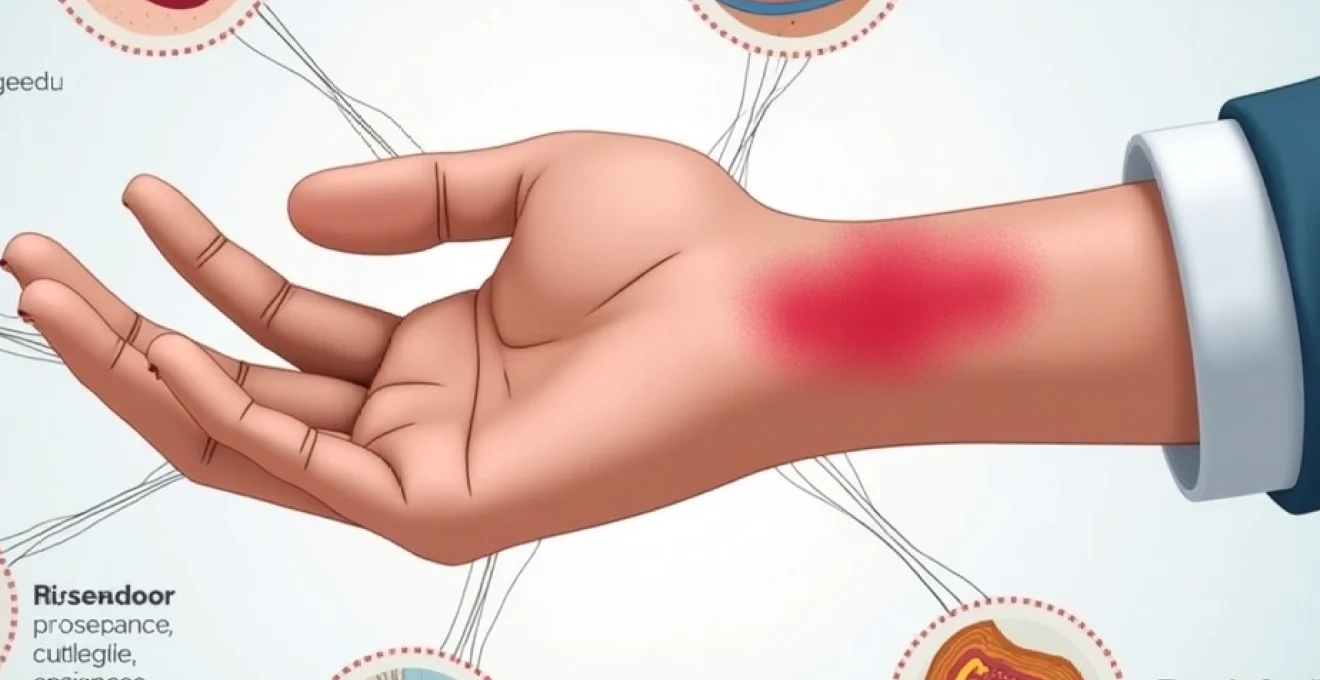
Haematoma formation and secondary inflammatory responses
Haematoma development following venipuncture represents a common complication that can trigger secondary inflammatory responses and cutaneous manifestations. These collections of extravasated blood occur in approximately 15-20% of blood collection procedures, with higher incidence rates observed in elderly patients, those taking anticoagulant medications, or individuals with bleeding disorders. The size and extent of haematoma formation directly correlate with the degree of subsequent inflammatory response and associated dermatological symptoms.
Subcutaneous blood extravasation pathophysiology
Blood extravasation into subcutaneous tissues occurs when venipuncture damages the vessel wall beyond the immediate puncture site, allowing blood to leak into surrounding tissues. This process is exacerbated by factors such as inadequate pressure application post-procedure, patient movement during needle insertion, or multiple puncture attempts at the same site. The volume of extravasated blood determines both the visible extent of bruising and the magnitude of the subsequent inflammatory response. Large haematomas can compress surrounding tissues, potentially causing nerve irritation or compromising local circulation.
Iron deposition and Haemosiderin-Induced skin discolouration
As extravasated blood undergoes degradation, the breakdown of haemoglobin releases iron that becomes deposited in tissues as haemosiderin. This iron deposition creates characteristic brownish discolouration that can persist for weeks or months following the initial injury. The process of haemoglobin catabolism also generates biliverdin and bilirubin, contributing to the typical colour progression of bruises from initial dark red or purple through green and yellow phases.
Understanding this natural progression helps differentiate normal healing processes from pathological complications that might require medical intervention.
Complement activation and cytokine release mechanisms
The presence of extravasated blood activates complement cascades and triggers the release of various inflammatory mediators, including interleukins, prostaglandins, and chemotactic factors. This inflammatory response serves to clear cellular debris and promote tissue repair but can also produce symptoms including pain, swelling, and erythema that extend beyond the immediate haematoma boundaries. Individual variation in inflammatory responses explains why some patients experience minimal symptoms while others develop significant discomfort and prolonged healing times following similar degrees of blood extravasation.
Pre-existing dermatological conditions and venipuncture complications
Patients with pre-existing skin conditions face increased risks of developing complications following venipuncture procedures. Conditions such as eczema, psoriasis, dermatitis herpetiformis, and chronic urticaria can significantly alter skin reactivity and healing responses. The compromised barrier function characteristic of many dermatological conditions increases susceptibility to both irritant and allergic reactions, whilst inflammatory skin diseases can be exacerbated by the additional trauma of needle insertion.
Eczematous conditions present particular challenges, as the already compromised skin barrier allows increased penetration of potential allergens and irritants. Patients with active atopic dermatitis show heightened reactivity to common phlebotomy materials, with reaction rates up to three times higher than in individuals with normal skin. The phenomenon of “angry back syndrome” can complicate patch testing in these patients, making definitive allergen identification challenging.
Autoimmune skin conditions such as pemphigus or bullous pemphigoid can be triggered or exacerbated by venipuncture trauma through the Koebner phenomenon. This isomorphic response involves the development of disease-specific lesions at sites of injury or trauma. Recognition of this potential complication is crucial for healthcare providers, as it may necessitate modified procedures or additional precautions in affected patients.
Clinical assessment and differential diagnosis of Post-Phlebotomy rashes
Accurate assessment of post-venipuncture cutaneous reactions requires systematic evaluation of multiple factors including timing of onset, morphological characteristics, distribution pattern, and associated symptoms. The differential diagnosis encompasses allergic reactions, irritant contact dermatitis, infections, haematoma-related inflammation, and exacerbation of pre-existing conditions. Healthcare providers must maintain high clinical suspicion for serious complications whilst avoiding unnecessary investigations for benign, self-limiting reactions.
The timing of symptom onset provides crucial diagnostic information, with immediate reactions (within minutes to hours) typically representing IgE-mediated allergic responses, whilst delayed onset (24-72 hours) suggests either T-cell mediated hypersensitivity or bacterial infection. Morphological assessment should document the size, shape, colour, and texture of affected areas, along with the presence of vesicles, pustules, or other specific lesion types that might indicate particular aetiologies.
Systematic questioning regarding exposure history, medication use, and previous reactions to medical procedures helps identify potential allergens or risk factors for complications. Patient reports of similar reactions to dental procedures, surgical interventions, or other medical contacts may reveal patterns of sensitivity to common healthcare materials. Documentation of current medications, particularly anticoagulants, immunosuppressive agents, or medications known to increase bleeding risk, provides context for assessing complication severity and healing expectations.