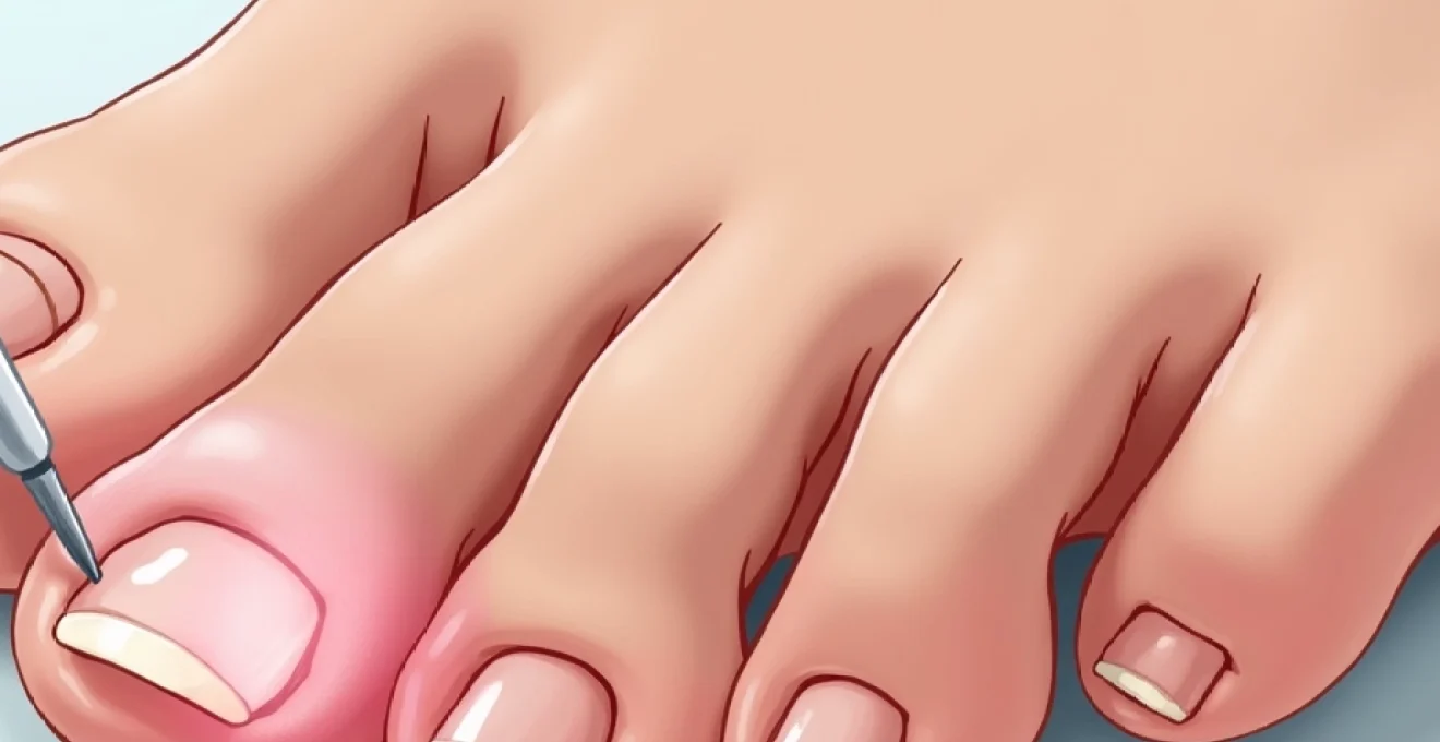
Losing a toenail can be both alarming and uncomfortable, leaving many wondering whether their nail will regenerate properly. The reassuring answer is that toenails do grow back in the vast majority of cases, though the process requires patience and proper care. Understanding the complex biology behind nail regeneration, the various causes of nail loss, and the factors that influence successful regrowth can help you navigate this common yet concerning situation with confidence.
The human nail apparatus represents one of the body’s most remarkable regenerative systems, capable of completely rebuilding lost tissue through an intricate process of cellular division and keratinisation. Whether your toenail detachment resulted from trauma, infection, or underlying medical conditions, the nail matrix—the growth centre located beneath your cuticle—typically retains its ability to produce new nail tissue. However, the timeline, appearance, and success of regrowth depend heavily on the underlying cause and the extent of damage to surrounding structures.
Nail matrix regeneration and toenail regrowth physiology
The nail matrix serves as the powerhouse of nail production, housing specialised cells called keratinocytes that continuously divide and differentiate to form the nail plate. This remarkable structure extends from the visible lunula—the pale crescent at your nail’s base—deep beneath the proximal nail fold, where it remains protected from external trauma. When a toenail falls off, the matrix typically remains intact and continues producing new nail cells, though the regeneration process follows a predictable yet gradual timeline.
Nail matrix cell division and keratinocyte production
Within the nail matrix, stem cells undergo rapid mitotic division, producing daughter cells that progressively differentiate into mature keratinocytes. These cells synthesise large quantities of keratin proteins, particularly the hard alpha-keratin that gives nails their characteristic strength and durability. The process of keratinisation involves the gradual flattening and hardening of these cells as they migrate towards the nail bed, forming distinct layers that eventually constitute the visible nail plate.
The rate of keratinocyte production varies significantly between individuals and can be influenced by factors such as age, nutrition, hormonal status, and overall health. Younger individuals typically experience faster nail regeneration due to more active cellular metabolism and enhanced blood supply to the nail apparatus. Temperature also plays a crucial role, with warmer conditions generally promoting increased cellular activity and faster growth rates.
Proximal nail fold protection during regeneration process
The proximal nail fold acts as a protective barrier, shielding the delicate nail matrix from mechanical trauma and bacterial contamination during the vulnerable regrowth period. This anatomical structure maintains the sterile environment necessary for proper keratinocyte function whilst providing structural support for the emerging nail plate. Damage to the proximal nail fold can significantly impact regeneration quality and may result in permanent nail deformities.
During the initial stages of regrowth, the proximal nail fold often appears swollen or inflamed as increased blood flow supports the heightened cellular activity beneath. This inflammatory response, whilst alarming to some patients, represents a normal physiological adaptation that facilitates optimal healing conditions. Proper wound care during this period helps maintain the integrity of these protective structures.
Nail bed vascularisation and growth factor distribution
The nail bed’s extensive vascular network plays a critical role in delivering essential nutrients and growth factors to support regeneration. This rich blood supply provides oxygen, amino acids, vitamins, and minerals necessary for keratin synthesis whilst simultaneously removing metabolic waste products. The characteristic pink colour of healthy nails reflects this abundant vascularisation, and changes in nail colour often indicate alterations in blood flow or oxygenation.
Various growth factors, including epidermal growth factor (EGF) and transforming growth factor-beta (TGF-β), orchestrate the complex cellular processes involved in nail regeneration. These signalling molecules regulate cell proliferation, differentiation, and migration, ensuring coordinated tissue reconstruction. Disruption of growth factor signalling can lead to aberrant nail formation or delayed healing, highlighting the importance of maintaining optimal systemic health during recovery.
Lunula formation and nail plate development timeline
The lunula’s appearance marks a significant milestone in the regeneration process, typically becoming visible within 4-6 weeks of nail loss. This pale, crescent-shaped area represents the distal portion of the nail matrix where newly formed nail cells begin their transition from soft, translucent tissue to the harder, more opaque nail plate. The lunula’s size and shape can provide valuable insights into the health and function of the underlying matrix.
As nail plate development progresses, the emerging tissue gradually extends beyond the lunula, covering the nail bed in a proximal-to-distal direction. The initial nail plate often appears thin and fragile, requiring careful protection to prevent re-injury. Complete structural maturation of the new nail plate can take several months , during which time the tissue gradually thickens and hardens to achieve its final mechanical properties.
Traumatic avulsion versus pathological toenail loss mechanisms
Understanding the underlying cause of toenail loss significantly impacts both the regeneration timeline and the likelihood of successful regrowth. Traumatic avulsion, the most common cause of nail detachment, typically results from acute mechanical forces that separate the nail plate from its underlying attachments. In contrast, pathological nail loss occurs gradually due to disease processes that compromise the structural integrity of the nail apparatus or disrupt the normal attachment mechanisms between the nail plate and nail bed.
Acute Trauma-Induced nail plate detachment patterns
Blunt force trauma to the toenail commonly occurs during sports activities, household accidents, or occupational injuries. The mechanism typically involves compression forces that cause subungual haematoma formation, with accumulated blood creating pressure that gradually separates the nail plate from the underlying bed. This process may occur immediately following severe trauma or develop gradually over several days as blood continues to accumulate in the subungual space.
The pattern of nail detachment following trauma can range from partial avulsion affecting only the distal nail plate to complete separation involving the entire nail structure. Partial avulsions often heal more successfully as the remaining attached portion provides a scaffold for regeneration whilst maintaining some degree of nail bed coverage. Complete avulsions require more extensive healing and carry a higher risk of complications such as infection or abnormal regrowth patterns.
Onycholysis secondary to fungal infections
Fungal nail infections represent one of the most common pathological causes of gradual nail separation and loss. These infections typically begin at the distal nail edge or lateral nail folds, progressively spreading proximally as fungal organisms colonise the nail plate and underlying tissues. The infection process produces enzymes that break down keratin proteins, weakening the nail structure and compromising its attachment to the nail bed.
Onychomycosis often presents as a slowly progressive condition characterised by nail thickening, discolouration, and brittleness before complete detachment occurs. The affected nail typically appears yellow, brown, or white, with a crumbly texture and foul odour. Fungal nail infections require specific antifungal treatment to prevent reinfection of the regenerating nail tissue and ensure successful long-term outcomes.
Psoriatic nail dystrophy and complete nail loss
Psoriasis affecting the nail apparatus can cause significant structural changes leading to nail plate separation and loss. The inflammatory processes associated with psoriatic nail disease disrupt normal keratinocyte function within the nail matrix, resulting in the production of abnormal nail tissue. Characteristic features include nail pitting, oil-drop discolouration, subungual hyperkeratosis, and onycholysis that may progress to complete nail loss in severe cases.
The autoimmune nature of psoriasis means that nail involvement often reflects the overall disease activity and may require systemic treatment to achieve optimal regeneration outcomes. Topical therapies alone are frequently insufficient for severe nail psoriasis, and biological treatments may be necessary to suppress the underlying inflammatory processes. Recovery from psoriatic nail loss often takes longer than traumatic cases due to the ongoing inflammatory environment.
Chemical burns and caustic substance nail damage
Exposure to strong acids, alkalis, or other caustic substances can cause severe damage to the nail apparatus, potentially affecting both the nail plate and the underlying matrix. Chemical burns often result in immediate tissue destruction, with the extent of damage depending on the concentration of the caustic agent, duration of exposure, and promptness of treatment. Such injuries may cause permanent damage to the nail matrix, potentially preventing successful regeneration.
The healing process following chemical nail injuries requires careful monitoring for signs of infection and assessment of matrix viability. Early intervention with appropriate wound care and neutralisation of the caustic agent can significantly improve outcomes and preserve regenerative capacity. However, severe chemical burns may require surgical intervention and carry a higher risk of permanent nail loss or deformity.
Clinical assessment of nail matrix viability
Determining the viability of the nail matrix following toenail loss requires careful clinical evaluation of both visible and palpable signs of healthy tissue function. Healthcare professionals typically assess matrix integrity through observation of the proximal nail fold, examination for signs of active bleeding or inflammation, and evaluation of the patient’s pain response in the affected area. The presence of a healthy blood supply, indicated by appropriate tissue colour and capillary refill, suggests good regenerative potential.
Advanced diagnostic techniques, including dermoscopy and high-resolution ultrasound, can provide detailed visualisation of the nail matrix structure and blood flow patterns. These tools help identify areas of permanent damage that may compromise regeneration whilst distinguishing between temporary inflammation and irreversible tissue destruction. Early accurate assessment of matrix viability guides treatment decisions and helps establish realistic expectations for regrowth outcomes.
The timeline for clinical assessment varies depending on the cause of nail loss, with traumatic cases typically showing clear signs of healing within 2-3 weeks. Pathological nail loss may require longer observation periods to distinguish between active disease processes and healing responses. Regular follow-up appointments allow for monitoring of regeneration progress and early identification of complications that may require additional intervention.
Toenail regrowth timeline and growth rate variables
Toenail regeneration follows a predictable but lengthy timeline, with complete regrowth typically requiring 12-18 months for most individuals. This extended timeframe reflects the slow growth rate of toenails, which advance approximately 1-2 millimetres per month under normal circumstances. The regrowth process occurs in distinct phases, beginning with matrix reactivation and early cell production, progressing through nail plate formation and emergence, and culminating in the restoration of normal nail thickness and appearance.
Multiple factors influence the rate of toenail regeneration, including age, overall health status, nutritional state, and local blood circulation. Older adults typically experience slower regeneration due to reduced cellular metabolism and decreased growth factor production. Systemic conditions such as diabetes, peripheral vascular disease, and autoimmune disorders can significantly impair the healing process and extend the regeneration timeline.
Environmental factors also play a crucial role in determining regrowth rates, with warmer temperatures generally promoting faster nail growth through increased metabolic activity. Seasonal variations in growth rates are well-documented, with peak regeneration typically occurring during summer months. Proper nutrition, particularly adequate protein intake and sufficient levels of biotin, zinc, and iron, supports optimal keratinocyte function and can positively influence regeneration speed.
The key to successful toenail regeneration lies in maintaining optimal conditions for cellular growth whilst protecting the vulnerable regenerating tissue from further injury or infection.
Individual variation in regeneration rates can be substantial, with some patients achieving complete regrowth within 10-12 months whilst others may require up to 24 months for full recovery. Factors such as the extent of initial damage, presence of underlying medical conditions, and adherence to protective measures during healing all contribute to these differences in outcomes.
Complications and aberrant nail regeneration patterns
Despite the nail apparatus’s remarkable regenerative capacity, various complications can arise during the regrowth process, potentially resulting in permanent structural abnormalities or functional impairments. These complications range from minor cosmetic concerns to significant problems that may require ongoing medical management or surgical intervention. Understanding the potential for aberrant regeneration helps patients maintain realistic expectations whilst recognising when professional intervention may be necessary.
Pterygium formation and nail bed scarring
Pterygium formation represents one of the most challenging complications of nail regeneration, occurring when scar tissue from the proximal nail fold extends onto the nail bed surface. This fibrous tissue can physically block normal nail growth patterns, resulting in permanent nail deformity or incomplete regeneration. The condition often develops following severe trauma or infection that damages the delicate anatomical boundaries between different nail structures.
Prevention of pterygium formation requires meticulous wound care during the early healing phase, with particular attention to maintaining the natural anatomy of the proximal nail fold. Early recognition and treatment of developing pterygium can prevent permanent deformity , though established cases may require surgical correction to restore normal nail growth patterns. The surgical approach typically involves careful excision of scar tissue and reconstruction of normal anatomical relationships.
Onychauxis and thickened nail plate development
Onychauxis, characterised by excessive nail plate thickness, commonly develops following traumatic nail loss or chronic infection. This condition results from hyperactivity of the nail matrix, producing abnormally large quantities of keratin that accumulate to form an unusually thick nail plate. The thickened nail often appears yellow or brown and may cause discomfort when wearing closed footwear.
Management of onychauxis requires regular professional nail care to maintain appropriate thickness and prevent complications such as ingrown nails or pressure ulceration. Mechanical debridement using specialised podiatric instruments can effectively reduce nail thickness, though the condition typically requires ongoing maintenance. Severe cases may benefit from chemical or surgical matrix reduction to permanently decrease the nail’s growth potential and prevent recurrent thickening.
Onychocryptosis risk following regrowth
The development of ingrown toenails (onychocryptosis) represents a common complication during nail regeneration, particularly affecting the great toe. Changes in nail shape, growth direction, or surrounding soft tissue relationships during healing can predispose to nail edge penetration into adjacent skin. The risk is heightened when the regenerating nail plate grows in a curved or pincer-like configuration that differs from the original nail shape.
Preventive measures include proper nail trimming techniques, appropriate footwear selection, and early intervention at the first signs of nail edge irritation. Professional guidance during the regrowth period helps establish optimal care routines that minimise ingrown nail risk whilst supporting healthy regeneration. Treatment of established ingrown nails may require minor surgical procedures to remove the offending nail portion and prevent recurrence.
Professional management and accelerated regeneration protocols
Professional medical management of toenail regeneration can significantly improve outcomes and reduce the risk of complications. Healthcare providers specialising in nail disorders employ various techniques to optimise healing conditions, monitor progress, and intervene early when problems arise. These interventions range from basic wound care and protective dressings to advanced therapies designed to enhance cellular regeneration and prevent aberrant healing patterns.
Modern regenerative medicine offers several promising approaches to accelerate nail regrowth, including the use of growth factors, stem cell therapies, and bioengineered scaffolds. Platelet-rich plasma (PRP) treatments have shown particular promise in enhancing nail matrix function and promoting faster, more complete regeneration. These advanced therapies work by concentrating natural healing factors at the site of injury, creating optimal conditions for cellular repair and growth.
Professional oversight during nail regeneration not only improves cosmetic outcomes but also significantly reduces the risk of long-term complications that could affect foot function and comfort.
Pharmaceutical interventions may include topical treatments to prevent infection, systemic medications to address underlying conditions affecting regeneration, and specialised preparations designed to enhance keratinocyte function. The selection of appropriate treatments depends on individual patient factors, including the cause of nail loss, overall health status, and specific risk factors for complications. Regular monitoring allows for adjustment of treatment protocols based on regeneration progress and early identification of potential problems.
Patient education plays a crucial role in successful professional management, encompassing proper wound care techniques, recognition of warning signs requiring immediate attention, and lifestyle modifications that support optimal healing. Collaborative care between patients and healthcare providers typically yields the best outcomes, combining professional expertise with consistent home care practices that maintain ideal conditions for regeneration throughout the lengthy healing process.